What should I do if the protein gets denatured during cryo sample prep? I’m using a 300-mesh carbon-coated gold grid, the particles at 200kV can be seen clearly but the 2D class averages don’t have high-resolution information.
1 Like
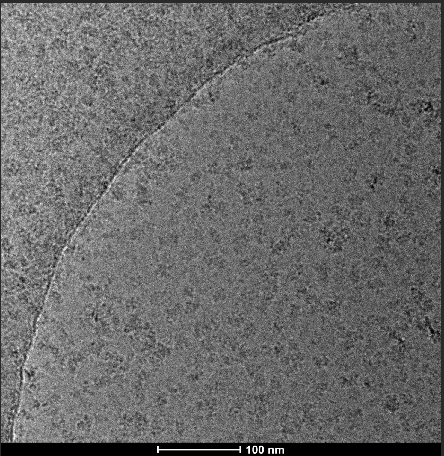
Hi Yovo, could you show the 2Ds? My first reaction when I glanced at the micrograph was “that looks great!” If the 2Ds have the expected particle shape I would not assume they’re denatured. If they’re lacking any hint of internal structure or the 3D reconstructions don’t go to hi res, something else might be causing that issue.
Hi drichman! This is my 2D classified images. I’ve adjusted the parameters many times, and this is what it looks like with relatively noticeable grain.
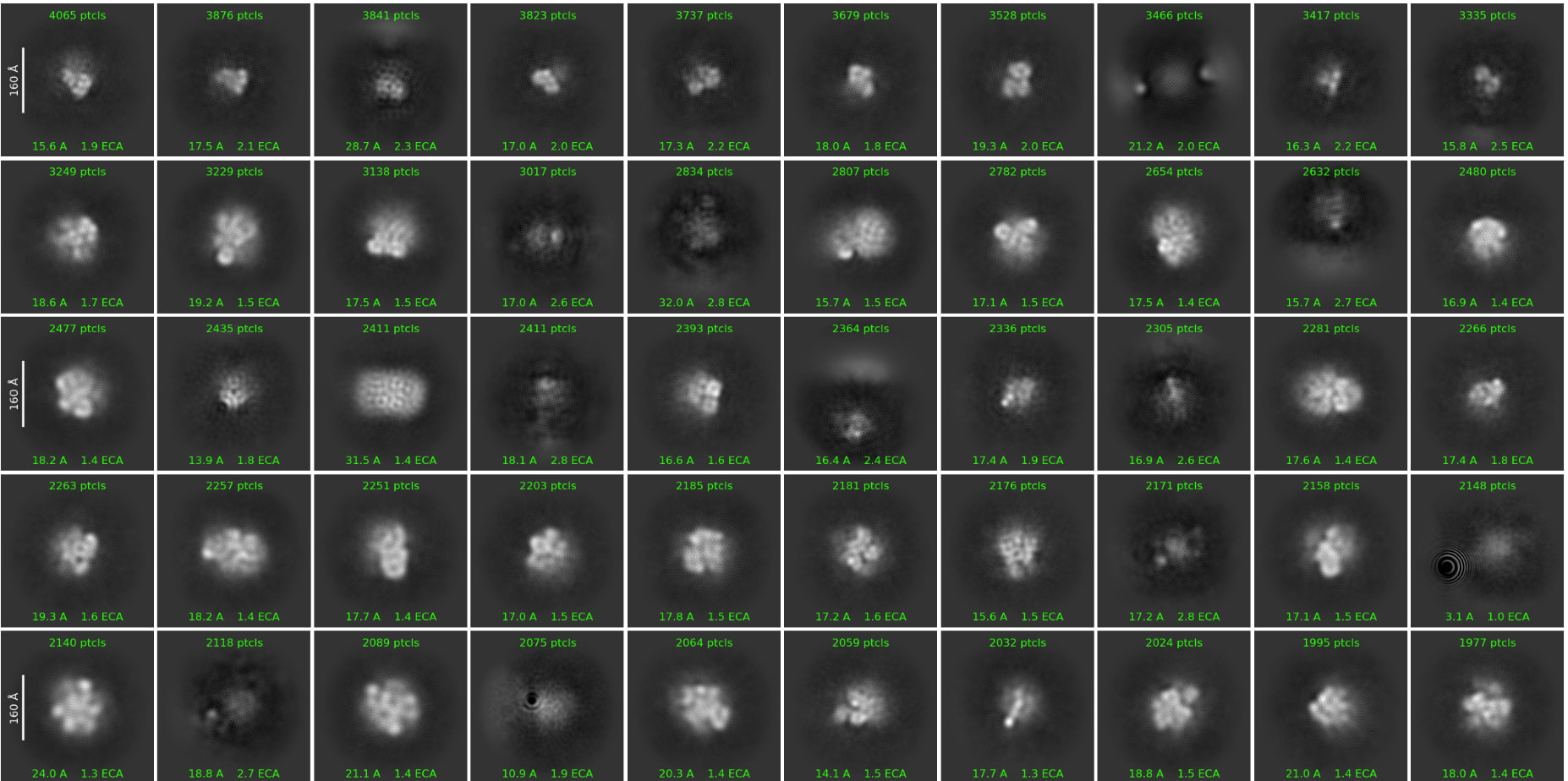
Ah I see. I guess those particles are not in as good shape as I thought at quick glance - assuming the particle picking makes sense by eye of course (centered on particles, not too much open area or junk etc).
Have you checked around the grid for other ice thickness? Sometimes the ice is too thin to support the properly folded protein. It’s also possible and very common that the protein doesn’t tolerate the air-water interface in open-hole grids, leading to denaturation. You could test detergents, other compounds that block the interface, grid surfaces (amorphous carbon, graphene oxide)… there’s range of techniques people have tried for this unfortunate problem.
Also double check if it’s not just a cryoEM problem - ie is the protein folded in negative stain conditions or its purification/bulk storage conditions.