Hi,
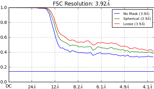
Recently, there are three data sets show abnormal FSC curve after the heterogeneous refinement like below. The FSC curve doesn’t go down to 0 and it is above the 0.143 line. Particles were extract from relion and imported into cryosparc. CTF estimation was done in relion using GCTF giving maximum resolution 4A. So i don’t know what is the reason? The original collected micrograph has problem? The GCTF parameter given is unsuitable? Or some other reason? Does anyone has similar problem?
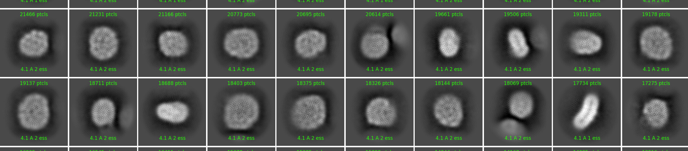
2D micrographs seems like with no problem, but i find the resolution reported of 2D classes is abnormal. Every class has the same reported resolution. Classes of Bin4 particles all show 7.1 A resolution. Classes of Bin2 particles all show 3.9 A resolution. Before these data sets, there is another datasets also extract from relion, but its 2D classes reported normal different resolution.
The FSC curves could be normal. Are your particles downscaled for the refinement? What were the pixel and box size upon extraction and what box size did you use for the refinement job? Note that the default value for heterogeneous refinement is 128 px. So, if your new pixel size is ~2Å and the data are good, it would be normal that the curve doesn’t drop to 0.
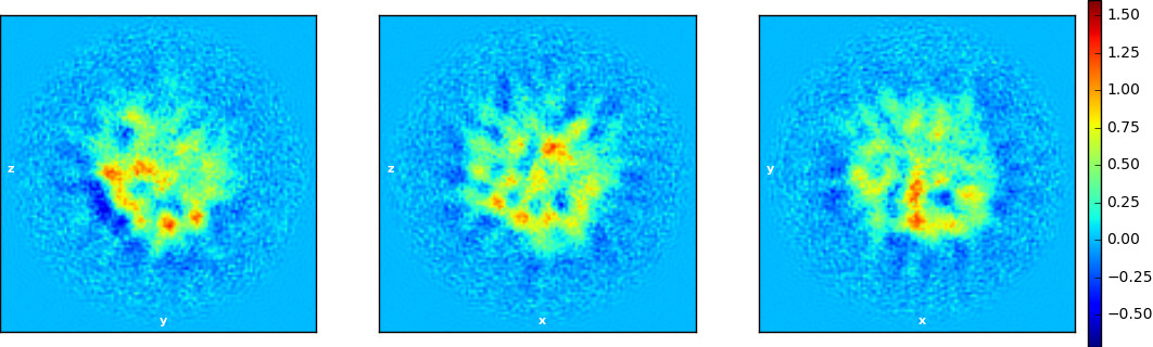
With that said, the real space slices don’t look great and I guess the volume presents artifactual stripes when you open it in chimera (?). Also, the 2D averages below present artifacts (these weird pentagonal “donuts” that make them look like a mosaic). We also have seen similar artifacts in my lab. I think the cause of this is failure to correctly align the particles, possibly due to huge variability, resulting in “overfitting”. It looks like you have a small membrane protein in a nanodisc (?). So the inner protein features are too little to guide the alignments and the outer scaffold protein is too variable (in conformation and size) resulting in these blurred averages. Good luck!
Probably others have more to say about this and I’m very curious to read what they have to say. In any case you should not trust the 2D resolution, at 4Å you should be able to clearly see secondary structure features, which is not the case.
This is usually due to merging of particle stacks that contain duplicates. Are you certain there are no duplicate particles? You can run the duplicate particle job to eliminate such particles if they all came from the same set of micrographs.
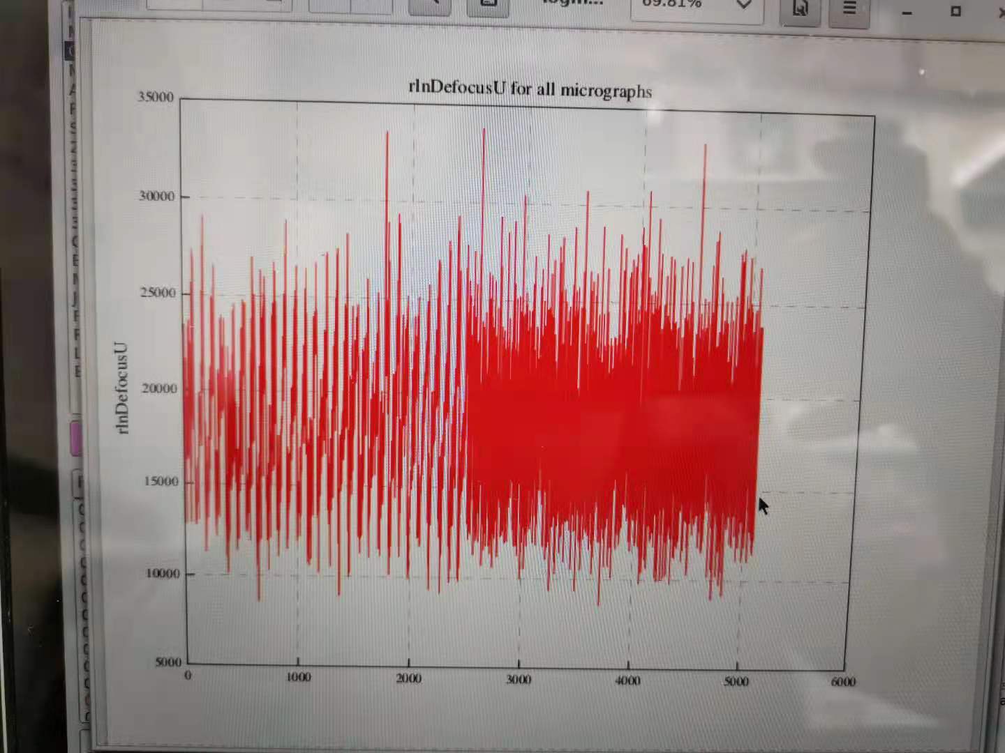
Thank you very much! The data set has some duplicated particles. But it may be not the reason. I found defocusU show abnormal pattern after CTF estimation. when i divide micrographs into two part. The problem was gone. But I don’t know why.
Hi! Thanks for your specific advice. The original pixel size of this data is 0.85A. Particle extraction was done using box size 288 rescaling to 72(bin4) or 144(bin2) for 2D classfication. In the 3D refinement, particles re-extracted with pixel size 0.85A and box size 288 was imported into cryosparc with default box size 128px. The small soluble domain is flexible. Protein is purify with detergent. It may actually has some overfitting. But in previous data of this protein. We didn’t meet this problem. Moreover, I found defocusU show abnormal pattern after CTF estimation. when i divide micrographs into two part, the problem was gone. But I don’t know why.