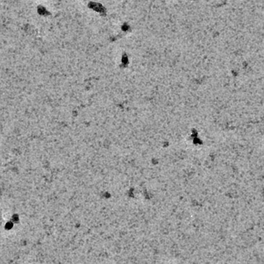
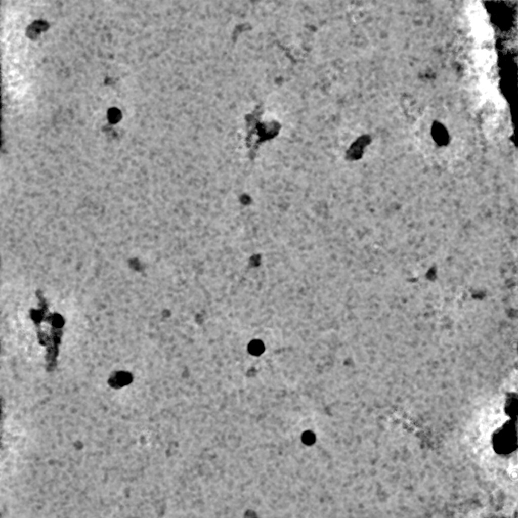
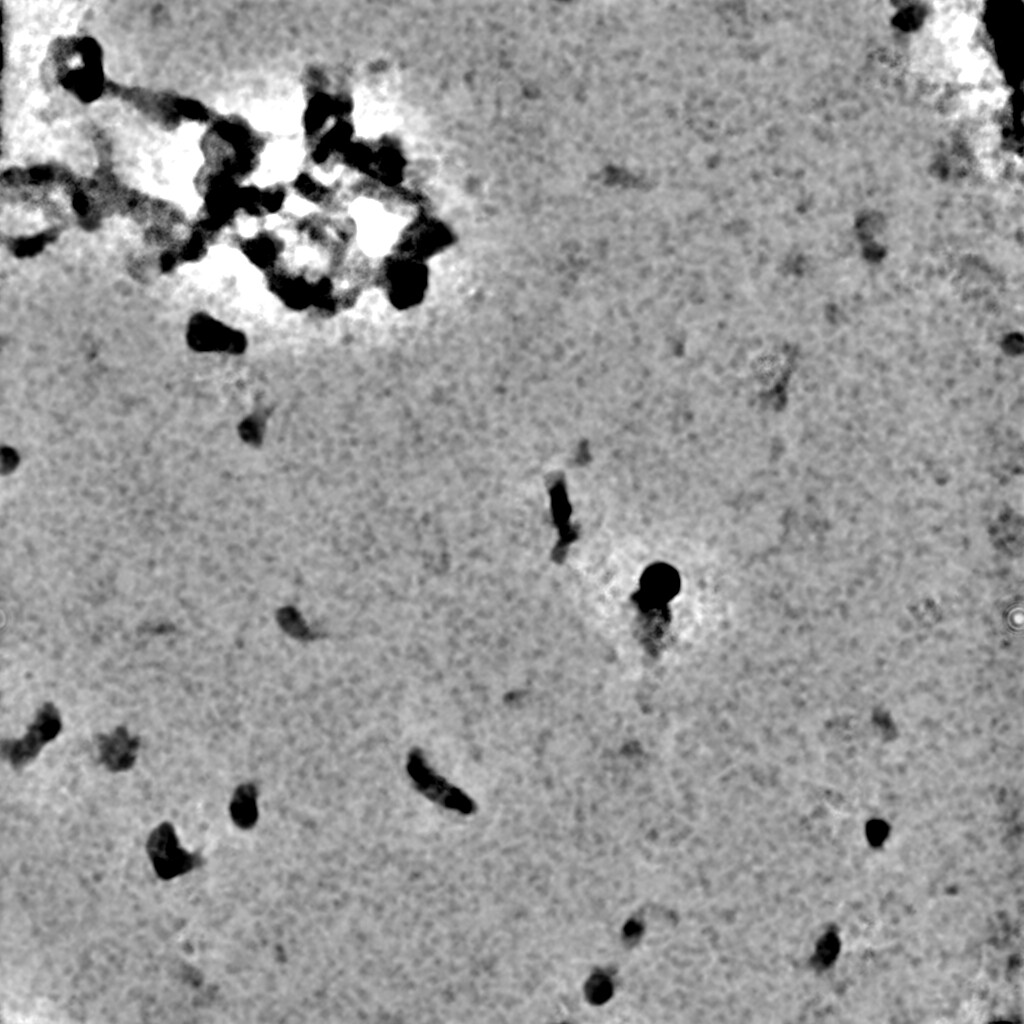
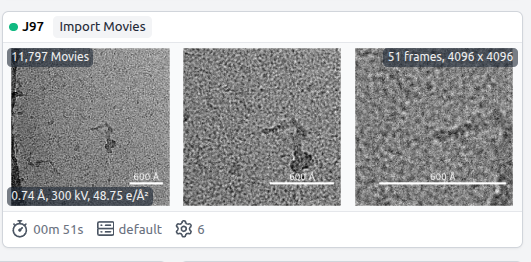
I have attached the denoised micrographs for reference. My particle size is approximately 100–105 Å. The data were collected at 165kX magnification with a pixel size of 0.74 Å.
I would like to know what box size should be used for the initial particle extraction and what would be the appropriate inner mask diameter for this dataset.
I have used 70-140 A blob diameter for particle picking.
Are you sure that is your sample, and that is isn’t broken? I see very little on those micrographs which are the appropriate size for a 10 nm diameter particle at your reported magnification…
Would you please highlight a few examples of what you think are your target of interest?
At 0.74 Å/pixel on a Falcon (likely on an energy filter as well given reported mag for pixel size) - which is what those micrographs look like they’re from - the field of view is ~300 nm. That means that a particle of ~100 Å (10 nm) should be approximately 1/30th the length of an edge.
Is there anything else about the sample which might explain the high background (carbon film used? detergents?) and did you use the pre-trained model (which gives poor results) or train the denoiser on your data (which can suffer if background is extreme)?
As a general point, if you’re trying to decide on optimal picking diameter and box size, the manual picker allows you to visualise and adjust both circular diameter (which can help educate potential parameters for blob picking) and box size (which allows you to choose an appropriate box size - ideally 2-2.5x the diameter of your particle, but higher symmetries can be analysed with smaller boxes without too much potential loss in resolution…
This is dimer protein, and yes it may exist in both the form. But protein which I have used for freezing, it was pass through size exclusion column and only dimeric population was incubated with small RNA molcule (25 nt) and then it was used. Whtever protein was remaining after grid freezing, I have done western blot and EMSA and both showed positive result.
This are quantifoil Cu grids 1.2/1.3 300 mesh size. For Denoiser job I didnt used pre-trained model. I used default settings for training.
Size of the particle I have mentioned is based on alphfold model of Protein+RNA and its quiet extended. Ideally it should be closed conformation. But I will mark few paricles.
This was also my impression also when I saw that micrograph. I would suspect that the sample has problems at air water interface. We have seen similar small damaged particles when we have had this problem in the past.
The high background and what appear to be very small particles relative to what you expect to see in the ice. This was what rbs_sci was point out. This is what makes me suspect that they may be damaged.
Thank you. Now I understood. Thank you for prompt reply. I have tried to pick the particles using 70-140 A blob. And I have extracted using 260 A box size bin to 180 A. I have started 2D classification using inner mask diameter of 100 A and outside diameter 130 A. I will update as soon as job get finished.
I see @jcoleman has been answering while I was busy, and following the same line of thought as I.
A few points, though:
“Field of view”: this is the edge-to-edge view of the micrograph - which would be 4096x0.74=3031Å (or ~300nm). Here:
You are describing the scale bar.
If your particles are broken, changing picking parameters will not help.
Running a dimer down SEC, seeing only a dimer and then incubating with something else does not mean it is still a dimer. Was the Western blot carried out with CN-PAGE, BN-PAGE or SDS-PAGE? If that latter, the presence of protein in the blot is meaningless for the presence of the complex.
Same with EMSA - all it means is that your protein interacts with DNA/RNA - not that your sample is actually in one piece.
Good you trained the denoiser on your data.
I presume from the lack of confirmation either way that, in fact, a carbon support film was not use?
Did you mark particles then remove the image? I cannot see an image with what you think are particle marked…
I have another dataset collected from graphene oxide grids. So I am transferring data to our cluster. I will denoise those micrograph and will check for the same and post it here with few marked particles.