I have been struggling to get good 2D classifications, in particular the side views of my interested membrane protein.
The membrane protein is a trimeric beta-barrel, stablised in detergent micelles. Trimeric size is 50x3=~150kDa, with a particle diameter of ~100A.
After reading multiple threads in the forum, particles were extracted with the box size of 250A (after motion correction and CTF find) for 2D classifications.
I got very good top-down view that clearly indicated the presence of the trimer (with three distinct holes). However, I could only get a blob with limited details which I assumed them to be side views and I want to improve this.
I’ve tried the following parameter settings for 2D classification:
I have also tried to select only 2 classes (guessing to be side views) of ~100k particles for 2D classification.
And they don’t look much like beta-barrel… I’m not sure if it’s due to the front and rear layer of beta-sheets crossing of the barrel that makes it so difficult to align?
Any idea what else to try? Any feedback would be helpful and appreciated!
Do you mind providing a micrograph for reference? What method did you use to pick the particles?
The 2D classification parameter settings you’ve tried seem reasonable enough (some suggestions follow later). What settings did you use to obtain the result you posted?
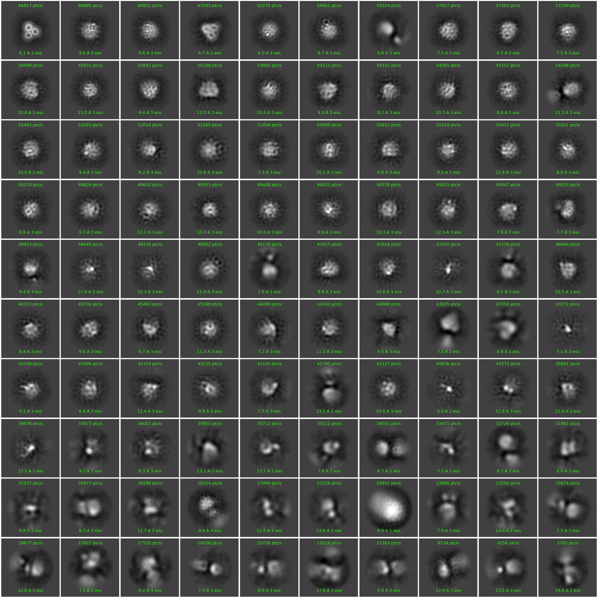
Could you provide how many particles you are classifying, and into how many respective classes? (Judging by the number of particles in each class average, i estimate over 1.5 million). In general with that amount, you would want to initially:
Classify these into 150-200 classes,
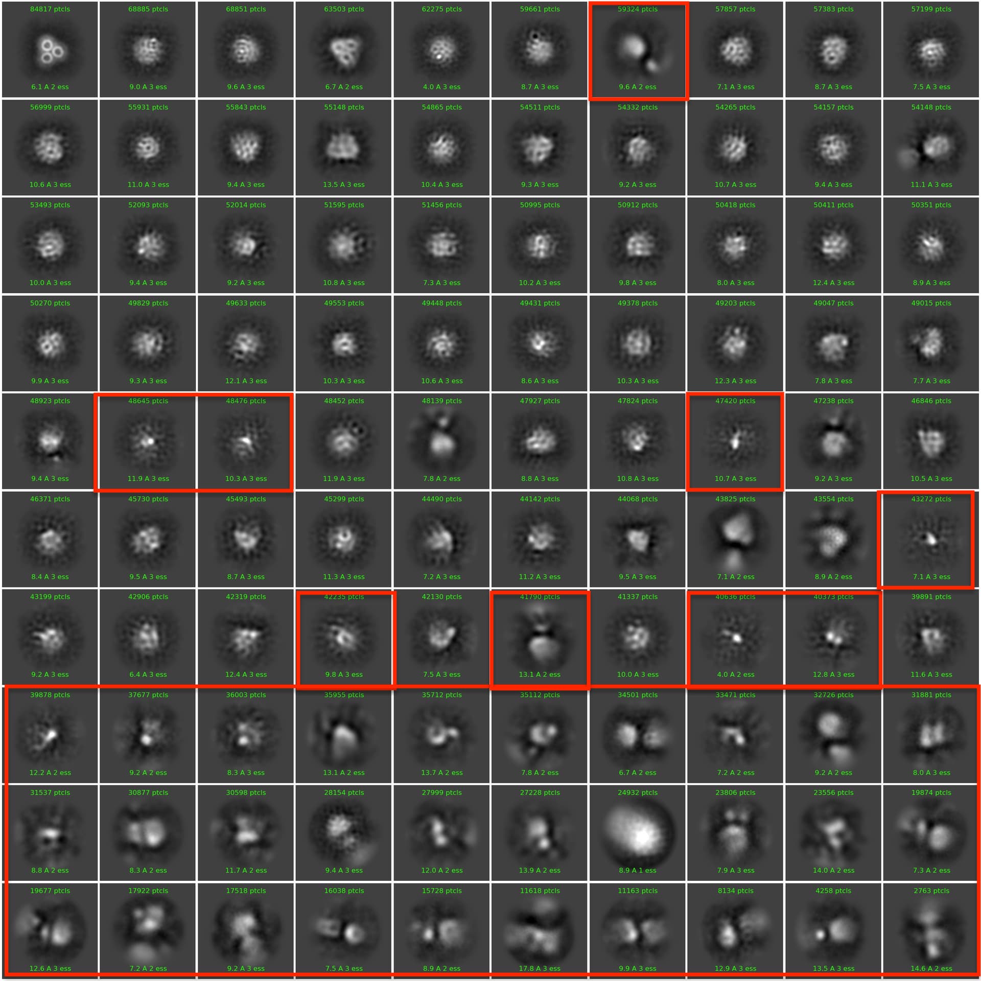
Exclude only obvious junk/crowded classes (see my attached image below. For example, exclude classes boxed in red)
You may start to tease out different views and better looking class averages by this iterative process, and could give Ab initio reconstruction a go with the cleaned up particle set. Probably don’t need to change re-center mask threshold in this case and best to try Force max over poses/shifts turned on as well and compare the results (i imagine you tried already).
Of course, the assumption here is that some side views are present in the data. It would be easy to imagine that top/bottom/oblique views could dominate due to air-water interface interaction/surface charge properties. Or these blurry classes are particles from relatively thicker ice areas/damaged particles/contaminant… Anyway, it is worth to try more extensive cleaning up of the data. Good luck and let us know how it goes!
I followed @ChaiG’s advice and did serial 2D classifications with 200 classes, force max over poses/shifts OFF, 40 online-EM iterations, and 400 batchsize. I left the rest of the parameters on default.
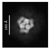
These are the only reasonable classes I’m getting so far, which look like a trimer to me:
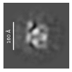
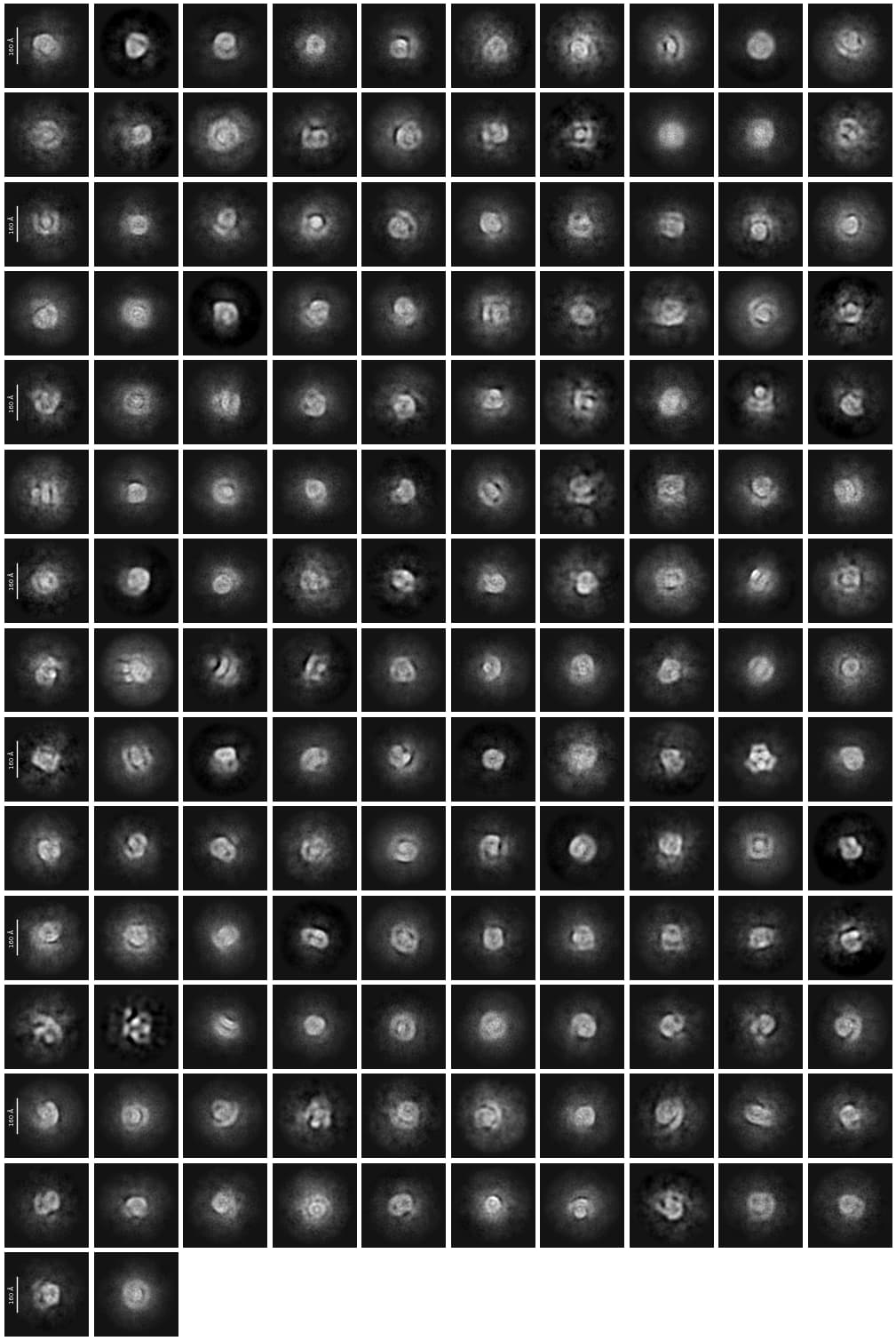
These are all of the classes, excluding the ones that are simply noise, which don’t look very nice:
Do you think there are protein micelles in my dataset, and that different views are not aligning for some reason? Or do you think these classes are something other than protein, for example, empty micelles?