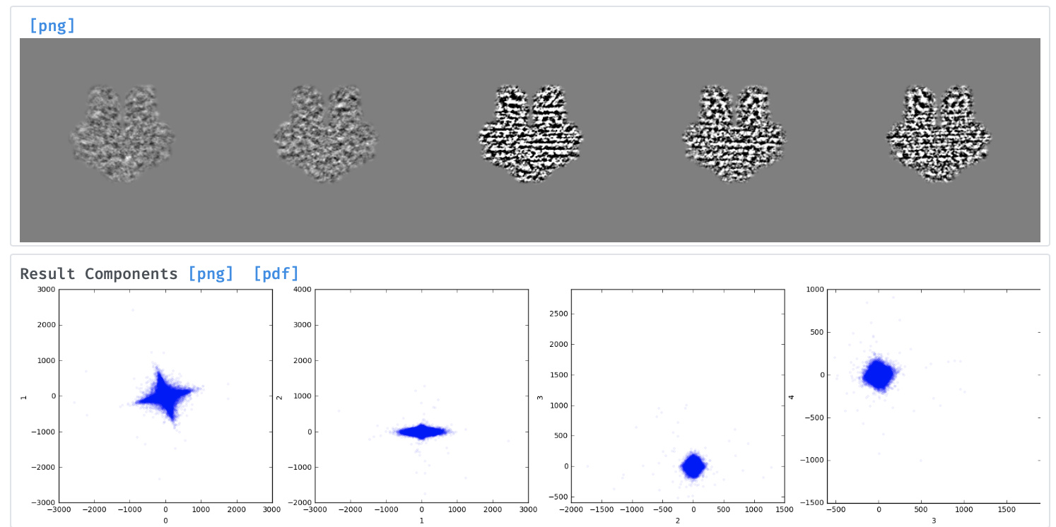
Although the frequency is much reduced in the latest version, I am still seeing some artefacts in the 3D variability analysis, marked by striping in the eigenvector images and characteristic “star-shaped” distributions in the reaction coordinate plots (see below for STRA6). Any suggestions on how to address much appreciated!
Hi @olibclarke the lambda parameter still controls the regularization that mitigates this issue and the default value (which is now normalized to values between 0-1) is only 0.01. I’ve also seen one case just yesterday where it needed to be turned up to 0.2… can you try that and let us know?
Cool - there shouldn’t be much negative consequence from increasing it though after a certain point the regularization will start to drown out the actual variability signal. The “streaking” seems to be related to having somewhat strong orientation bias - it’s the directions of the missing views that end up developing streaking.
PS with the STRA6 you may want to mask out the micelle entirely since there’s a huge amount of variability in it, that dominates the first many components before you actually get to seeing the protein moving around. In most membrane protein cases I’ve played with so far micelle masking is necessary - and the variations in the micelle don’t seem to correlate at all with changes in the protein density itself.
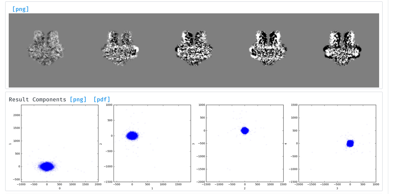
This is with the micelle (or nanodisc in this case) masked out actually… I wondered if it was related to orientation bias, because the “side view” of the mode image looks much weaker/fainter.
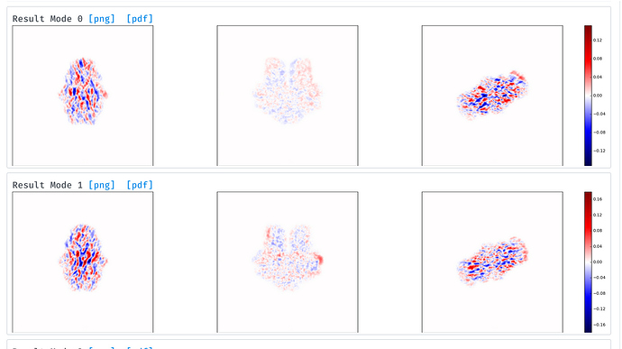
The mode images are not slices but rather projections of the mode along each axis - so even if the mode has a lot going on, if the total +/- density sums to zero along an axis, it’ll look washed out as you see here. The other projections do show that something is going on though so it can only really be interpreted in 3D!
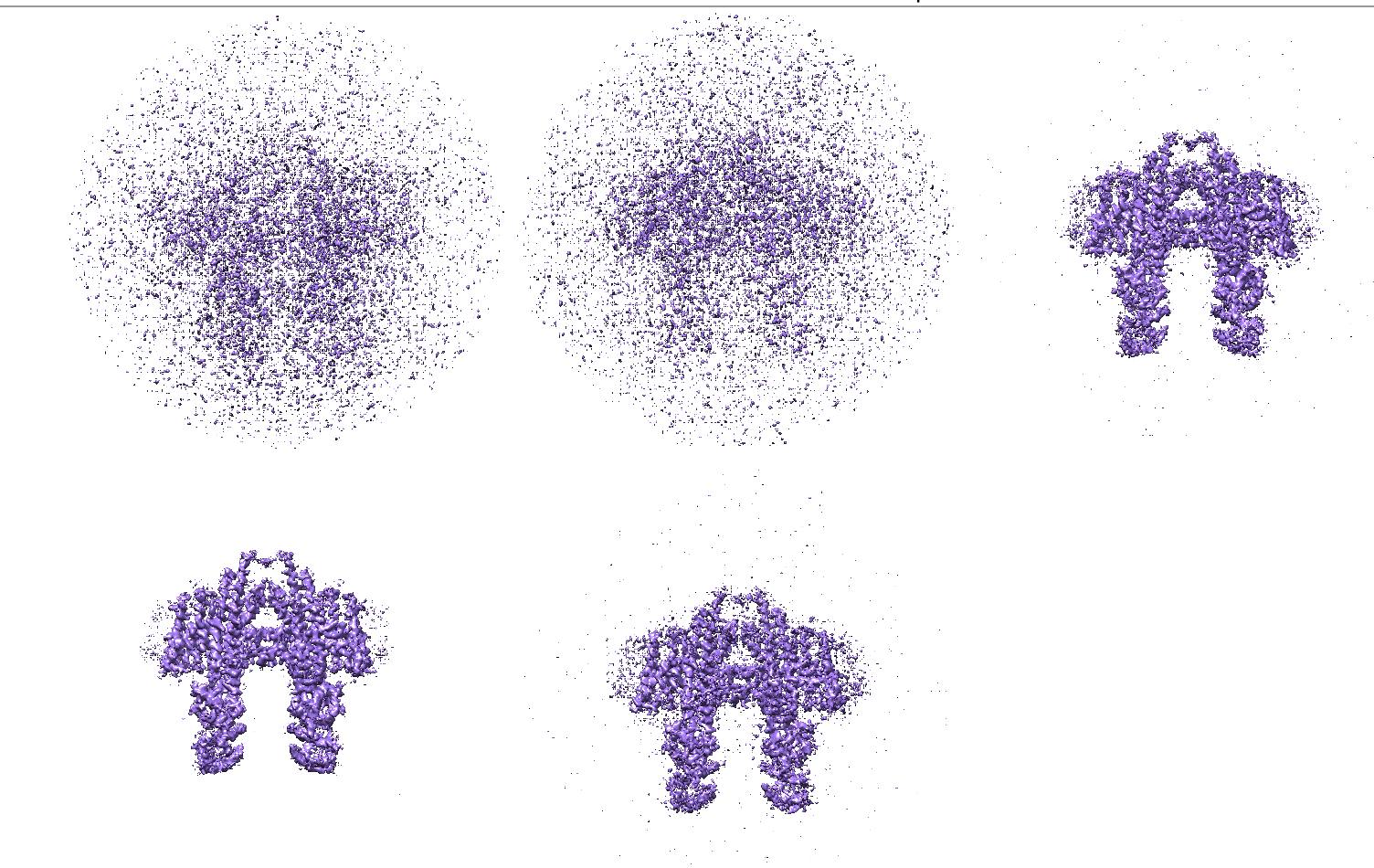
Yes, I looked in 3D, and it looks weird - it falls apart into noise as one progresses along e.g. mode 0. For the modes where something is going on (movement of the “legs”), that is still visible, but in the presence of ever increasing noise. Maybe a higher lambda will help.
I have this problem also as Oli mentioned in the case of membrane protein with micelles.
In my case, this artifacts was not caused by mask, instead it is caused by highpass filter (highpass resolution). I set the Highpass resolution to 20 as suggested in the 3DVA tutorial. As long as I have the highpass resolution set, I will have similar artifacts as Oli mentioned. Any update on solving this problem?@apunjani
@ZHEN do you only see artefacts when the highpass filter is set? If you leave the highpass filter to blank/None, do you still see streaking?
Can you also try with the noise model set to white (Use white noise model set to True)?
Thanks for help.
Yes, I only see artifacts when the highpass filter is set. I didn’t see streaking if leave the highpass filter to black/None.
I tried with noise model set to white but still got the streaking artifacts.