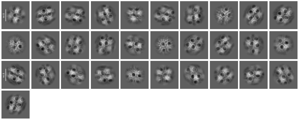
Recently I met a strange issue. As shown in the attached figure, during the processing of 2D-classification, we clearly observed some circle-shaped densities with a diameter of 3-4 nm, which somehow shaded our target protein, resulting in failure in aligning our target particles. Our sample is capable to attach with membrance fraction, so we first treated it with the detergent DDM and then removed the DDM by exchanging buffer for cryo-EM sample freezing.
Dose anyone meet this similar observation? What are the circle-shaped densities, some unknown binding proteins, remaining membrance fraction, detergent micelles or some artifacts due to misaligning? Any idea is welcoming!
What do your micrographs and individual particles look like? Are the individual particles the size that you are expecting (you can measure in ImageJ)? If you do inspect picks, does it seem like the particles are clearly circled and the circles are the correct size? If you are on gold grids, are you avoiding picking on the gold foil (or using junk detector to not pick on the gold foil)?
Based on looking at this, aside from the circles, it seems that each class may have multiple particles in the image (or just total noise) and this definitely increases your chance of getting weird artifacts – although I haven’t seen this flavor particularly, and I don’t work with membrane proteins much. You can try changing the 2D windowing to smaller if your box size is the classic ~2x particle size. You could try other tricks for small proteins (search past forum posts) to allow for better alignment as well.