Hello,
I’ve been using helical reconstruction on a fairly routine filamentous protein from our lab. I’ve previously collected data from a local microscope and using helical refinement with maximal symmetry 8, got this FSC curve:
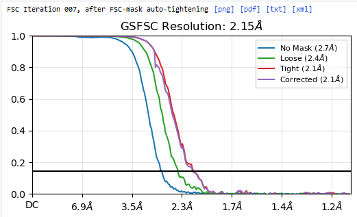
The FSC curve reaches 0 like expected.
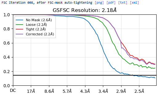
However using the same grids I collected at a different microscope with a slightly higher pixel size(0.537 instead of 0.54), processed the data in an identical way, and when I attempt to use helix refine with maximal symmetry 8 I get FSC curves like this:
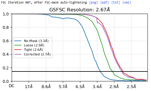
However when I repeat the process with maximal symmetry not set, the FSC curve returns to normal like so:
So clearly applying symmetry is causing a duplicate particle affect on the FSC, but why is it suddenly happening now?
Thanks for any insight.