Dear all,
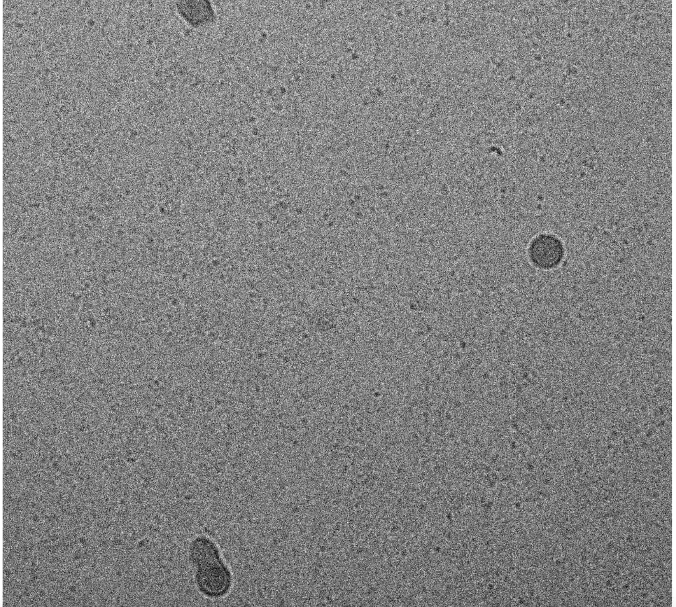
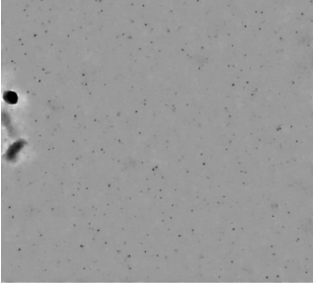
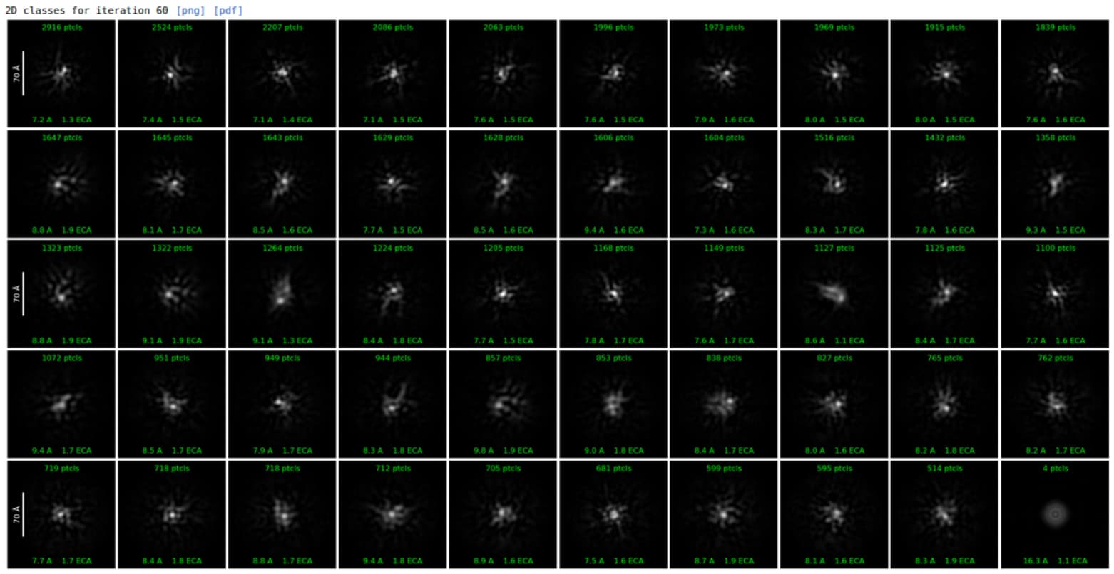
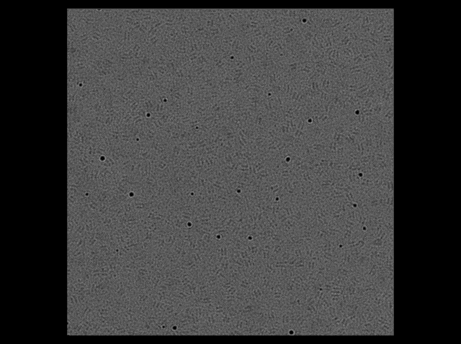
I have collected a dataset of gold-protein (71 kDa) using Falcon 4i as detector.I can see particles in micrograph, but couldn’t able to make good 2D except bright white spot inside the box.
Data collection: 300 kV, 50 e/A2 and 0.74 A pixel size, 8764 M
After preprocessing steps (Import movies/Patch Motion Correction/Patch CTF/Micrograph Denoiser/Inspect Particle Picks/Extract Micrograph), I went for 2D.
Is the gold necessary? Does you protein bind gold, or is it being used as a tag (in e.g., single molecule studies)? Because gold is very heavy and influences the electrons which interact with it much more strongly than carbon, nitrogen or oxygen (or phosphorus, for that matter). Even iron or zinc can cause issues (I can think of only two papers which have successfully reconstructed ferritin with iron present…) so I think with gold present you might struggle.
nice micrographs, the dark spots have a lot of contrast and remind me of others I had with some gold contamination (from the grid). It seems not all of the particles have gold here.
I would look into that papers from @rbs_sci, really interesting studies.
The more contrast could be mean more interactions with electrons, something in the buffer, cubic ice, etc. X-ray fluorescence and other methods with electrons could be useful for elemental analysis, but that seems like over kill.
TOPAZ could be trained to not pick the light particles, or just use the blob picker and bring the level down to a point where it does not select the dark ones - maybe a set where you bring it up to select only the dark ones. I think you can work with it, the denoised ones more dramatic and the training could misrepresent some of the low contrast particles. Now that I look closer I don’t think this is the correct micrograph + denoised, the large dark parts do not match.
Well, yes, because every atom in the beam path potentially effects the beam. Heavier atoms have more impact, which is why we use negative staining for example…
@SDcryoEM can you do the local refine with the other mask and make a composite map in Phenix ? You can be creative with the composite maps, but the real statistics only come from the local/focus refined map.