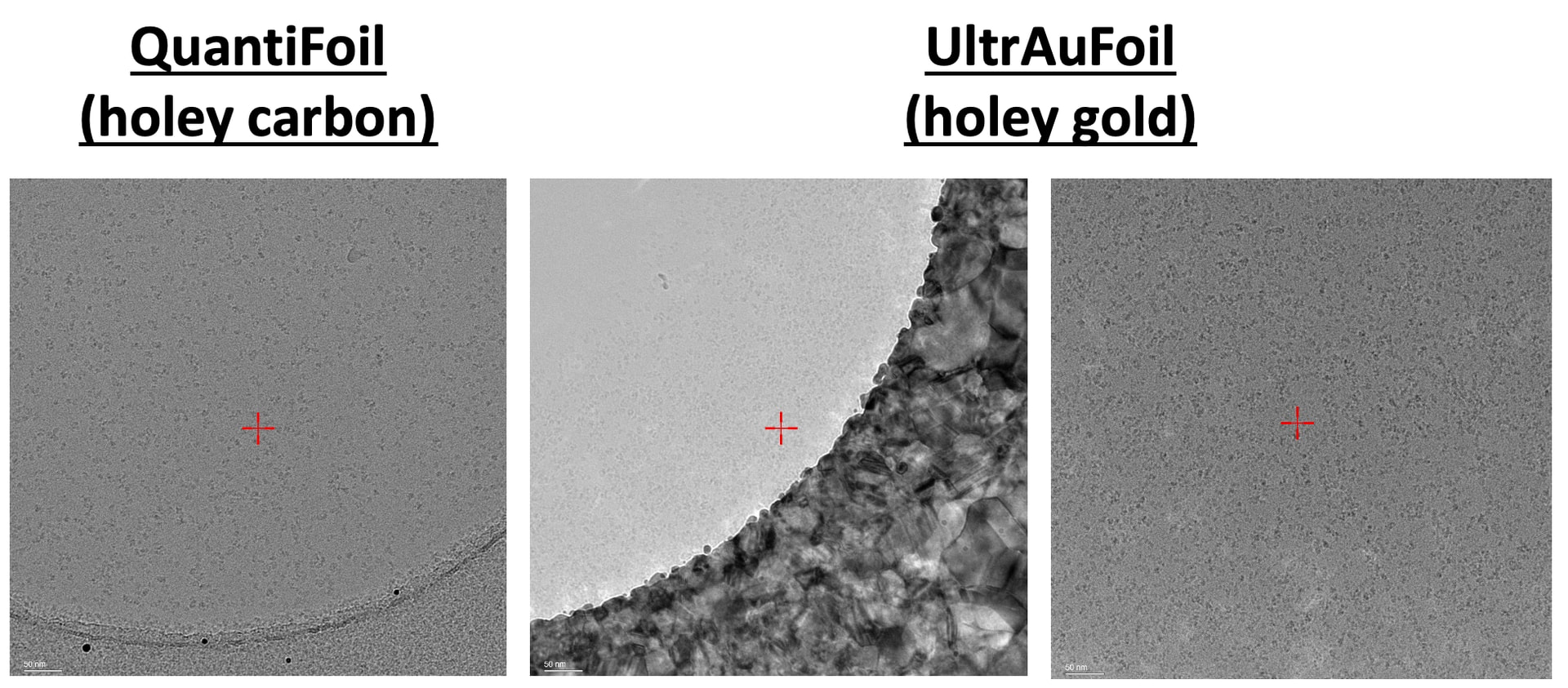
I recently prepared some grids using both QuantiFoil 1.2/1.3 holey carbon grids and UltrAuFoil 1.2/1.3 holey gold grids. While screening these grids, I noticed that with UltrAuFoil, whenever I image with the hole edges peeking inside the image, I get ultra-high contrast images where particles look nearly invisible. Typically, I shoot with hole edges (for holey carbon grids) because this helps a lot with CTF fits and improves throughput (using AFIS, I get more shots per hole if I shoot off-center).
Is the high-contrast in UltrAuFoil due to electrons being scattered from the gold support? Should I keep this in mind when I collect data (not just screening) on this UltrAuFoil grid? If so, do I need to keep both my imaging square/beam circle inside of the grid circle with ice and avoid the edges? I’m attaching a QuantiFoil grid screening image as comparison. Thanks for your help! Looking forward to giving these new grids a try and processing the data. I am currently planning on collecting data on a Krios that will hopefully give everyone here an idea of these grids’ performances: first day will be spent collecting on UltrAuFoil, and the other day will be spent collecting on Quantifoil. Hoping to make the best use of my time and not accidentally mess up the UltrAuFoil collection… (Also, somewhat tangentially, would appreciate any thoughts on which grid to prioritize — what are your thoughts on the particle dispersion/ice quality between the two grids for example? This is a 150 kDa protein complex. Thanks!)
That’s normal with gold grids.
Different grid materials require different optimisations. What works well for copper is sub-optimal for gold (and vice versa). 
If you’re shooting that much carbon on sensor to acquire multiple shots per hole, if you have fringe-free-imaging, I’d recommend spending some time optimising beam size vs. dose to tighten the beam up so you can shift the sensor area further onto ice. 
Gold grids are hard/impossible to get good Thon rings on for astigmatism/coma alignment, and basically it’s the same issue when gold is caught in the sensor area for an exposure. Try to avoid catching gold as much as possible.
In your examples, the particle dispersion looks better on the AuFoil, so assuming a good orientation distribution I’d prioritise that, aiming at ice-only.
I’m still debating, given the cost and supply constraints of AuFoil relative to copper grids here, whether or not they are worth general usage. We struggled with a sample recently where we moved to gold grids and did see an improvement in resolution compared to copper with the same prep, but then with a new prep we blew straight through the gold results with copper. Previous tests have had them pretty much at a wash with optimal samples - 1.22Ang with gold, 1.24Ang with copper for apoferritin, 1.65Ang with copper and 1.75Ang with gold for a membrane protein complex. Some samples really need gold grids, others… 
Others will have further thoughts. 
1 Like
Very helpful to know all this — thank you! You must have some secret sauce if you all are pushing sub-2 angstrom.
I do plan to bring a “trash grid” for astigmatism/coma alignment. When you say “avoid catching gold as much as possible,” do you mean just avoid the sensor area (i.e. image box), and it doesn’t matter if the beam/illumination area touches the gold edges? What exactly causes this high contrast, and how does it interfere with particle information inside the ice? Apologies for my naïveté; this might be really obvious.