I am working on a 150 kDa membrane protein in a very tight nanodisc system. The scaffold protein is only 10A away from the membrane protein. NU refinement gives about 5.5 A resolution. Local resolution estimation shows that the scaffold protein is worse than 7A (there is density but no protein feature) whereas the membrane protein has ~4.5 resolution.
I hope to focus only on the membrane protein to further improve the alignment/resolution.
I have tried:
Varying the parameters in NU refinement: FSC threshold/Adaptive window factor
Create a mask then local refinement
Particle subtraction then local refinement
It is very difficult to create a mask that cleanly exclude the scaffold protein signal.
I wonder if there are other parameters in NU refinement that I can experiment with to tackle this situation.
Or is there a better processing strategy for this case?
Any suggestion is welcome. Thanks!
Don’t discount further cleanup of your dataset as a means to improve resolution. Either multi class ab initio, or heterogeneous refinement with one good model and several junk decoys, can often help improve resolution in such cases. You’d be surprised at the number of empty nanodiscs that hang around, in particular, even if you think you’ve removed them all.
Your suggestion worked!
Multi class ab initio couldn’t separate bad particles in my case.
I have done 5 rounds of heterogeneous refinement (1 good model + 2 junk models), followed by NU refinement on good particles. I have cleaned up my dataset from 130k to 60k particles.
The resolution from NU refinement went up from the original 5.5 to currently 4.6 A. The density of the membrane protein is now much more significant than that of scaffold protein and I started to see more feature of the membrane protein.
I wonder how many rounds do I have to do. I assume I will just have to keep going until the map isn’t improving and starting to become worse.
glad to hear it! Yep keep going until things start getting worse. You may want to vary the decoy models you use, too - sometimes I find it helps to generate decoys by running ab initio on discarded particles (to generate data-driven junk models)
Also once the resolution improves a bit, you may want to revisit local refinement (with or without signal subtraction) - it often gives a considerable improvement in such cases.
I could’t push the resolution further. I suspect the reason is too thick ice. I got CTF fit for my 9000 micrographs in average 5.5 Å, which is quite bad.
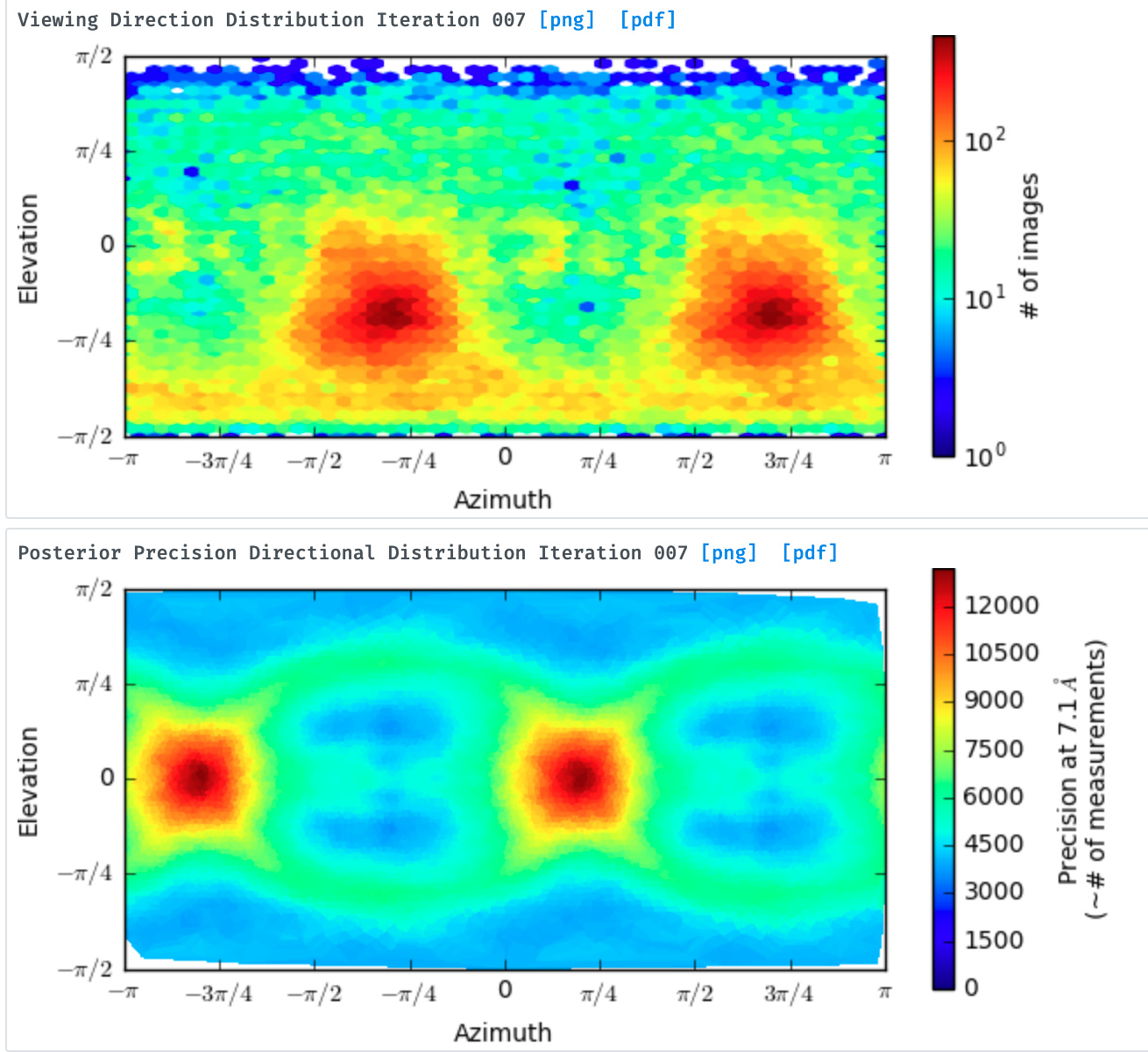
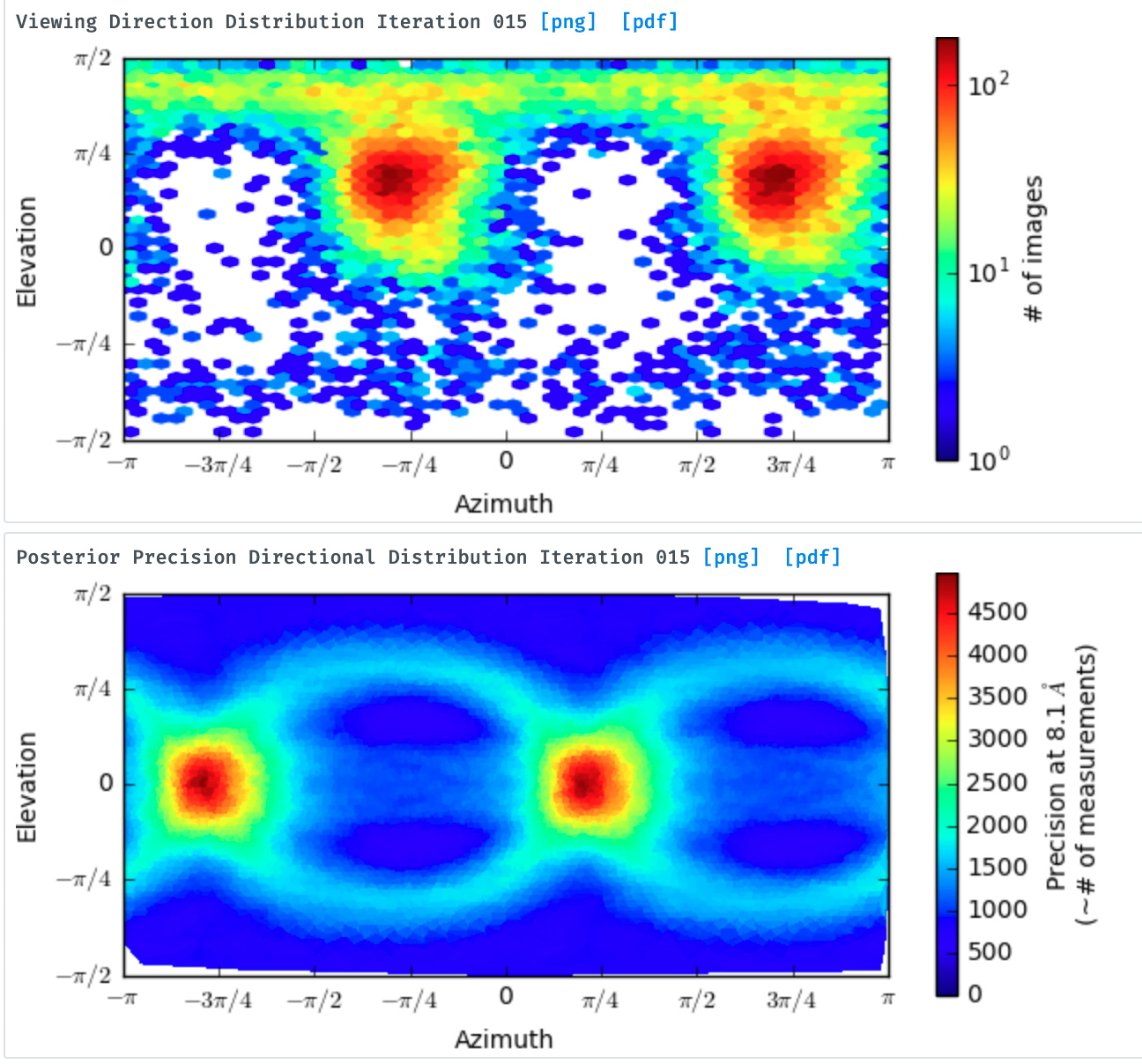
I probably also have a mild preferred orientation issue. Before I took @olibclarke suggestion, I got the viewing distribution like below:
I assume those views that were discarded are not good quality particles or those views are not even exist in my dataset. But the reconstruction actually looks okay (no elongated artefact). Could anyone provide their experience about viewing distribution, is this bad?
I am also experiencing problems with a membrane protein with a tight fit in a nanodisc. It seems that after ab initio reconstruction I can see the an obvious scaffold protein with central density but as soon as I try any refinement of the particles I get very little definition of any disc or central density. The refinements result mostly in noise. I have tried to make a mask around the central density to exclude the scaffold seen in the ab initio reconstruction but it makes no difference.
Is this a problem exclusively with my particle quality which I suspect is not good or am I doing something wrong? I think there is a fair amount of movement of the nanodisc relative to the membrane protein which I think it making this a very tricky sample. In addition I currently have some orientation bias in my sample. I am preparing more grids also but if anyone has anymore tips for how to work with the data I have and any future data that would be much appreciated.
I don’t believe there is any intrinsic issue with tight nanodisks or micelles. There are many published high-res structures in amphipol and tight micelles or NDs (TRPM4 in MSP1D1 for example is pretty extreme). I think general prep quality/homogeneity and/or particle size are most likely explanations.
I am also confused when you say the disk is both very tight and that there is substantial movement of the membrane protein.
 Yep keep going until things start getting worse. You may want to vary the decoy models you use, too - sometimes I find it helps to generate decoys by running ab initio on discarded particles (to generate data-driven junk models)