Occasionally I get mics with strange-looking ice (see attached), but it varies from hole to hole in the same square, and in many cases within the same image. This makes me suspicious that it is a temperature/freezing-related issue. I was wondering if others have observed this and/or know what may be causing it. Also, is there any simple way to remove these mics in manual curating aside from selecting them individually? Or will they be taken care of in 2D/3D. The particles are still clearly visible in most cases. Thanks.
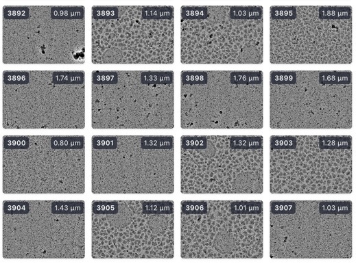
Yup, sometimes called leopard ice. As you say, usually temperature/freezing related. Can be indicative of poor contact between the grid and the holder. If you have any exotic additives in your buffer (organic solvents) that can also make ice more vulnerable to this happening as they can dramatically change the vitrification characteristics of the buffer…
1 Like
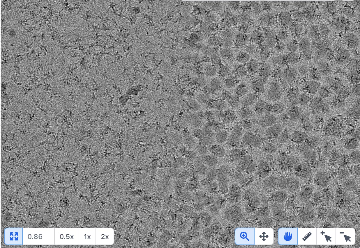
Are the particles extracted from leopard ice expected to be sorted/removable in 2D/3D pruning steps? If not, does anyone know of a machine learning algorithm to train and sort images with ice artifacts? Thanks.
You might try MicAssess (High-Throughput Cryo-EM Enabled by User-Free Preprocessing Routines - ScienceDirect) or Cinderella (cinderella_micrographs []) - I haven’t tried either so not sure whether they will help, but it sounds like the kind of scenario they ought to be useful for