Hi,
I’m new to the structural biology world. I have been processing the data for HIV Env (420kDa). This data set was collected on Tundra, 100kV, Pixel Size: 0.76A
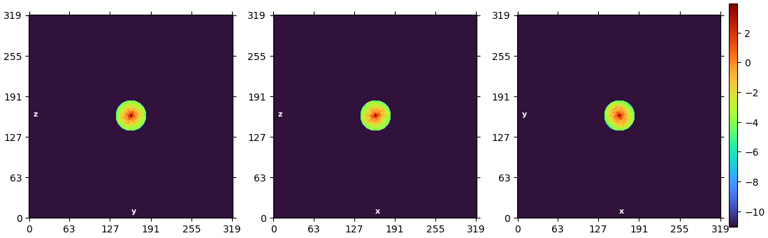
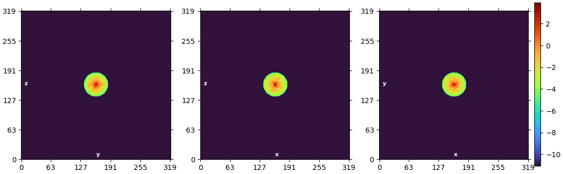
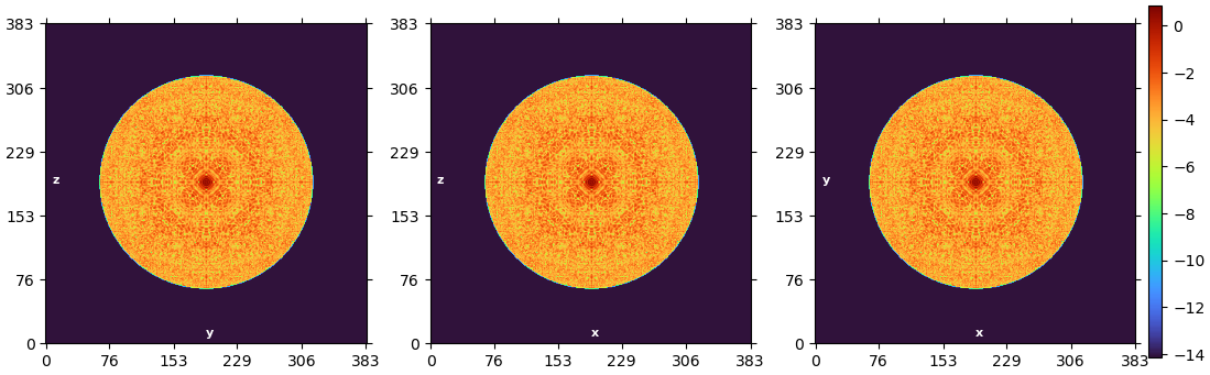
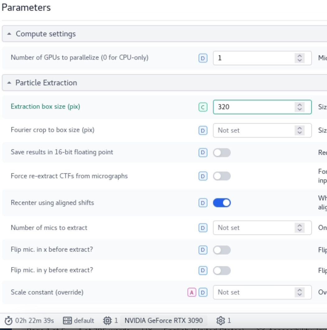
Initially, I have used extraction box size as 320pix and 80 pix as Fourier crop to box size. I got 9.85 A resolution after NU-refinement. Below is the image for the fourier space slices from NU-refinement.
If you Fourier cropped your particle images to 80 pixels for the first refinement, we would expect those boxes to only go from 0 to 79 — it looks like both refinements here used the full-size images. \
There are a few ways this might have happened. If you share the parameters you used for extraction we could tell for sure, but the most likely way I can think of is if you set the Second (small) F-crop box size parameter but used the full-size images. Alternately, if you used the small arrows in the text box to set the parameter, it may not have been set properly.
If you’d like to figure out why the particle images were not sucessfully F-cropped, please post the parameters you used in your particle extraction job and I can help you take a look .
As for why the Fourier circles look small in these images, that is expected if both of the refinements only go to low resolution.
CryoSPARC only uses low-resolution information during early iterations (set by Initial lowpass resolution) and increases them as/if the GS-FSC improves. In your case, the refinements are going to about 10 Å, and the edge of the box represents information with a frequency of approximately 1.5 Å. Thus, the information used in your map is only from a small circle in the center of Fourier space.
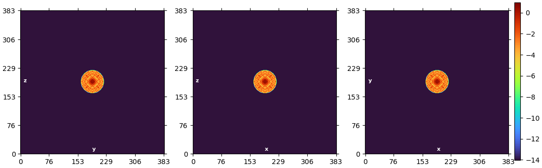
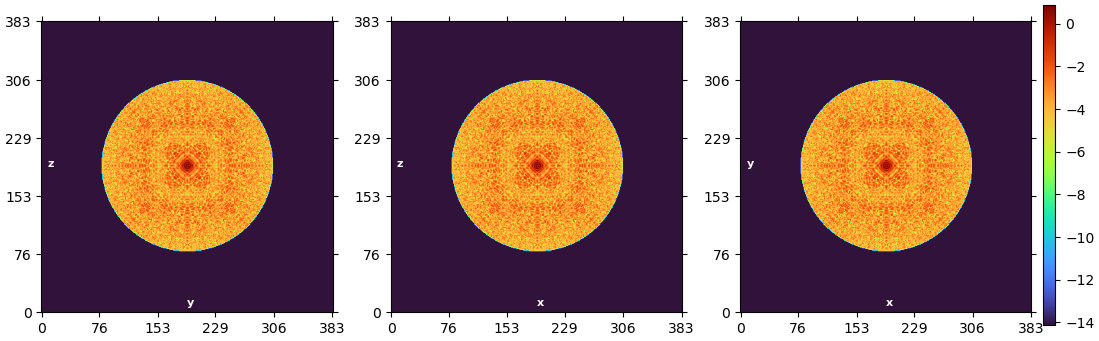
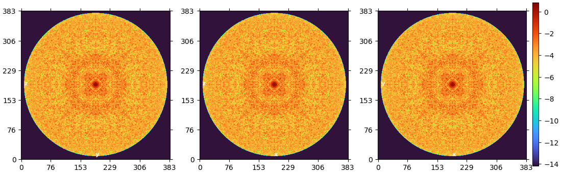
For comparison, here are these plots from the various iterations of a refinement that went almost to Nyquist:
As you can see, as the quality of the reconstruction improves, we use information from higher and higher frequencies, so the slice gets larger and larger.
I hope that clarifies things! Please let me know if there’s anything that’s still not clear!
These parameters worked very well for the data collected on Krios (300kV, 1.08 A Pix size). If I were to translate the same setting for data collected on Tundra (100kV, Pixel Size: 0.76A), how to get the right box size?
Hi @ml705! It looks like the Fourier crop to box size (pix) setting was left unset — this explains why both sets of images were 320 px.
As far as translating the data to the new dataset, since the pixel size is slightly smaller you will likely want a proportionally larger extraction box, assuming that both datasets are the same particle.
Your original box size was 320 px * 1.08 Å/px = 346 Å across, so the same box in your second dataset would be 346 Å / 0.76 Å/px = 455 px. When you’re picking an extraction box size, it’s important to pick a computationally fast box size — typically I’d pick the next box size that is larger than the one we just calculated (480 px in this case), but 448 px is pretty close, so that might work too.
I would definitely recommend significantly downsampling/F-cropping these particles to start. Since you’re only going to about 9 Å right now, the high-frequency information is not being used anyway, and downsampling the particles will significantly speed your processing.
Trying an extraction with box size 448 but Fourier-cropped to 128 would leave you with a final pixel size of 448 px * 0.76 Å/pix / 128 px = 2.66 Å/px. This means your downsampled Nyquist would be 2 * 2.66 Å = 5.32 Å, which would leave you plenty of room for improvement while significantly speeding up your analysis.
Of course, box size is just one of the many parameters you might need to change for new datasets. Parameters that work well on one dataset may not work well on another — part of the fun and frustration of SPA is how much exploration and “try-it-and-see” you have to do! So there’s no guarantee that these settings will improve the results, but they are likely a step in the right direction!