Hi all,
I need some insight for the interpretation of nu-refinement results.
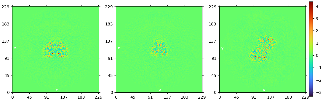
What does represent this color panel ? The protein I study exposed a C2 symmetry, here with a 230 pix box. During the first steps of processing, this panel was blue with a C1 symmetry and a 160 pix box. No mask was used for this refinement.
Thanks for your help!
Kevin