I am working on a small (~75 kDa) membrane protein with a soluble domain, solubilized in DDM (could be a 70-100 Å protein). The sample appears monodisperse and well-behaved based on cryo-EM screening. The estimated particle size in DDM is ~80 Å.
I picked ~400k particles and ran 2D classification in CryoSPARC with the following parameters:
Image acquisition: 105,000x, pixel size: 0.83 Å
Dose: 40 e⁻/Ų
Number of 2D classes: 80
Maximum resolution: 6 Å
Force max over-shift: off
Number of iterations: 80
Batch size per class: 400
Circular mask diameter: 120 Å
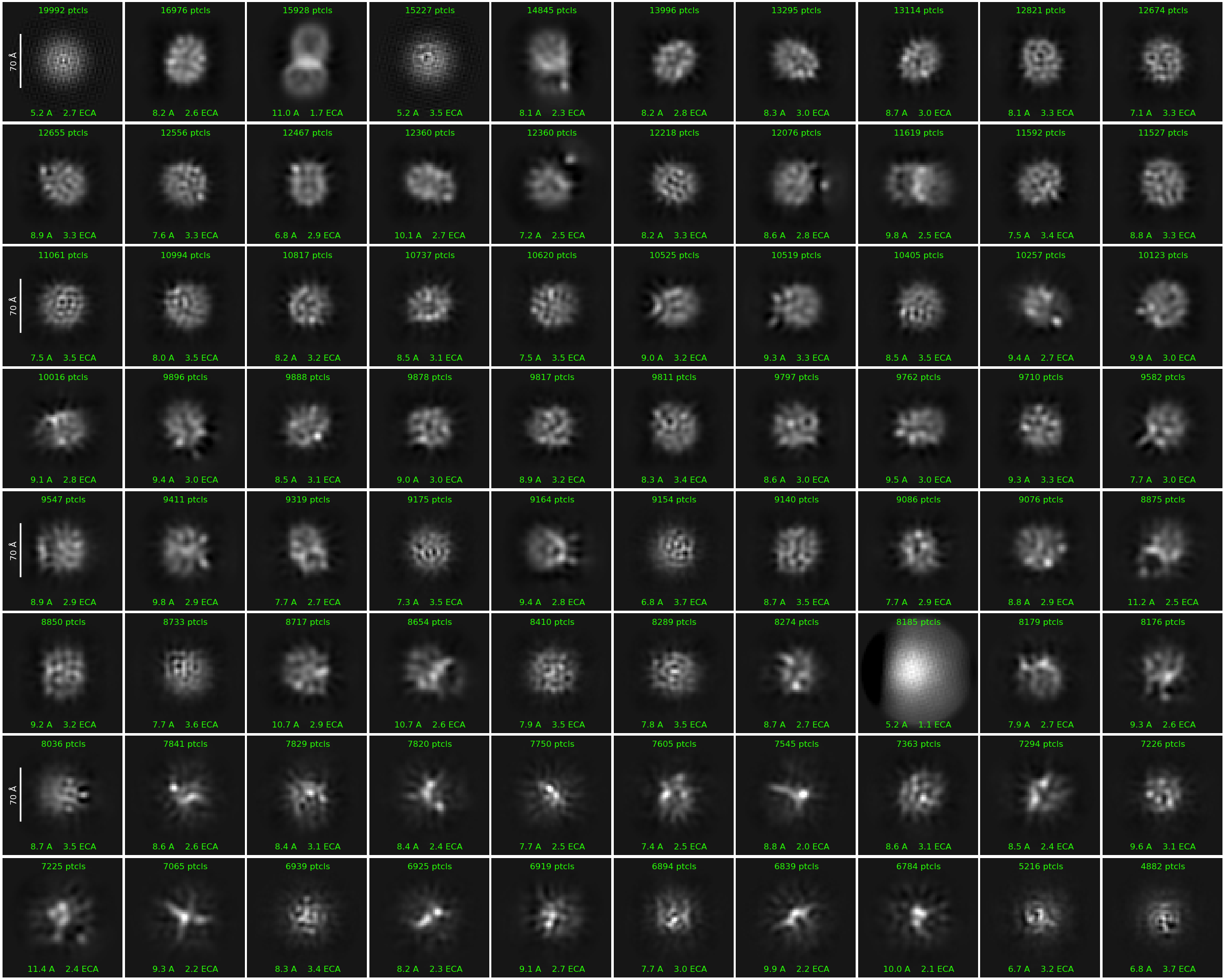
Although I can sometimes see a small soluble domain attached to the membrane domain, most of the resulting 2D classes do not show clear structural features.
I have also tried the denoiser tool, but the results were essentially the same.
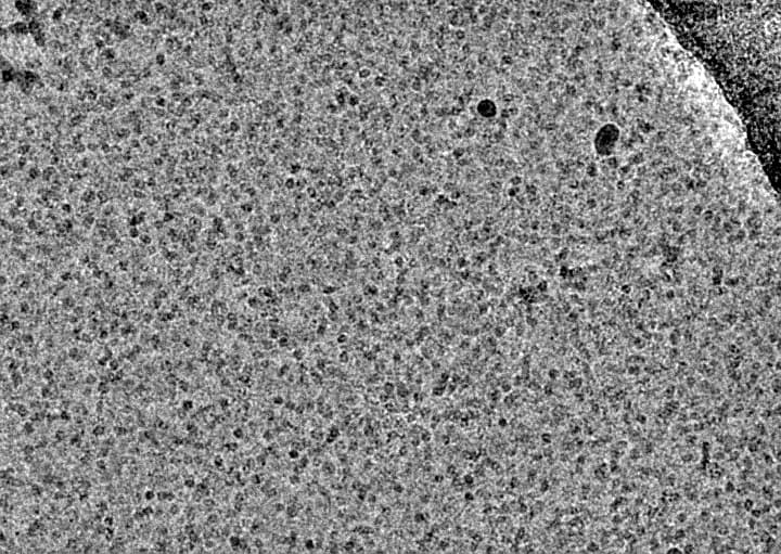
I have attached a representative micrograph and the obtained 2D classes for reference.
I would appreciate any suggestions on how to improve class quality. Could this be due to detergent effects, particle alignment, or classification settings?
Working with DDM is particularly critical for small membrane proteins. Make sure your protein concentration is high enough to maintain a strong signal alongside the DDM micelles. For a ~75 kDa membrane protein, 2 mg/mL is a good starting point.
Avoid concentrating your GPC peak more than 2–3x ; going higher also concentrates the DDM, which leads to the problem you’re seeing. The 2D classes suggest that most of what you’re picking up are empty DDM micelles.
Your processing parameters look fine, try increasing the number of classes to around 200 and see if that helps capture more meaningful particles.
I have screened various sample concentrations, and 2 mg/mL looked a bit too concentrated for this protein, so I reduced it to 1.6 mg/mL in the current preparation.
Okay, I will keep that in mind for my next prep (no more than 3x) and also try generating 200 2D classes to see if that improves particle separation and class quality.
Protein concentration actually to me seems on the low side to me which makes me think that your data may have been recorded on thicker ice. What is your average CTF fit resolution? We try to aim for better than 3 Ans on average. Collecting on minimally thin ice is critical for small membrane proteins.
We routinely concentrate some of our proteins to 6-9 mg/ml if they can tolerate this. I would not worry so much about concentrating detergent if your SEC peak fractions are around 0.5 mg/ml or better. Just concentrate through 100kDa cutoff. You will get some empty detergent micelles which you should be able to classify out. The key is to get as many particles as possible in each micrograph without aggregation in the thinnest ice possible.
Most of the CTF fits are well below 3.5 Å, with about 70% of the micrographs meeting that criterion. During particle extraction, I filtered for micrographs with CTF fits better than 5 Å to ensure data quality.
Moreover, for this preparation, I used a 100 kDa cutoff filter during concentration. I agree that ice thickness may still be a factor, especially given the relatively low protein concentration. I will look into optimizing ice thickness and possibly increasing the concentration further, depending on sample stability.
Looking at your 2Ds and micrograph, it looks like there are a lot of free micelles here. Agree re shooting for high concentration and thin ice. I also think your box size here is a bit small - I would suggest a box size of ~320px as a starting point (still using a 120Å mask for 2D).
Re this:
I have also tried the denoiser tool, but the results were essentially the same.
You may already be aware of this, but just to make clear - the denoiser will affect the micrographs that are used for picking, but particles are not actually extracted from the denoised mics. The denoising just helps visual interpretation and picking accuracy/precision.
a tip to not over-concentrate DDM after the SEC if tu centrifuge at 1000g instead of 5000g. It decreases the shear forces and decreases the non-newtonian behavior of the detergent mixture. Run 10 minutes at 1000g, mix then centrifuge again. 100kDa cutoff might be way to high for you, you’re lucky if its works but you might want to use 50kDa cutoffs, which definitely concentrates DDM (on regenerated cellulose; PES membranes might not).
Agreed that for this size protein, you might have to go to higher protein concentration.
DDM micelle size is around 70 kDa, this is roughly additive with the protein mass so the true size of the particle is around 140 kDa. I would use 100 kDa cutoff unless you have a good reason not to. Definitely agree that you should concentrate slowly and mix often.
FYI the detergent belt around a membrane protein is not the addition of the micelle + protein.
The aggregation number of DDM is betwen 80-120 depending on sources, while for a 70KDa membrane protein (I’m guessing 6-7 TM) there is roughly 200 DDM : visit the detbelt server for more visual ideas of what quantified amount of detergents look like around membrane proteins.
So the belt would be 200x510g/mol = 100kDa which is in theory big enough for the 100kDa concentrator. But, with centrifugation forces, the membrane dilates and the pores open up, up to twice the size (cf manufacturers). Also, the geometry of your protein has an influence.
Overall, a small membrane protein definitely can go through concentrators of 100kDa; I’ve personally experienced several times, with even 130kDa protein going through 100kDa pores… But I guess it’s protein dependent.
We will always try with a 100kDa cutoff first. Most proteins that are 50kDa or even smaller we find will not go through in DDM, MNG, or GDN micelles, this is to avoid detergent concentration as much as possible. We have a few rare cases where we have seen them going through but in that case, we adjust to 50kDa cutoff.
Looking at a representative reconstruction of a 68kDa protein that is basically just TMD without any soluble domains in DDM that was concentrated through a 100kDa cutoff, the measurements of such a particle is ~80 x 70 x 50 Ans. The smallest dimension of the thickness of the detergent you would think could be an issue but we generally don’t find it to be. This is just our experience.
Thank you for the helpful suggestions!
I agree that the high number of free micelles is something I have been worried about, and I will aim for higher concentration and thinner ice in my next prep. I will also try increasing the box size to ~320 px as you suggested while keeping the 120 Å mask for 2D classification.
One thing I am still unsure about is how to differentiate empty micelles from micelles containing protein just by looking at the micrographs. Are there any specific features that can help distinguish between them?
Thank you for the helpful discussion.
In my case, I ran a few trials using both 50 kDa and 100 kDa cutoff filters, and I agree that the output can vary from protein to protein. Luckily, I was able to retain most of the protein using the 100 kDa filter, therefore I am not worried about continuing to use it. However, I expected it to remove empty micelles, it was a bit surprising to see that it also seems to concentrate empty detergent micelles.
Sure, I will try concentrating the sample at 1,000xg for 10 minutes to see whether that helps improve the sample quality.