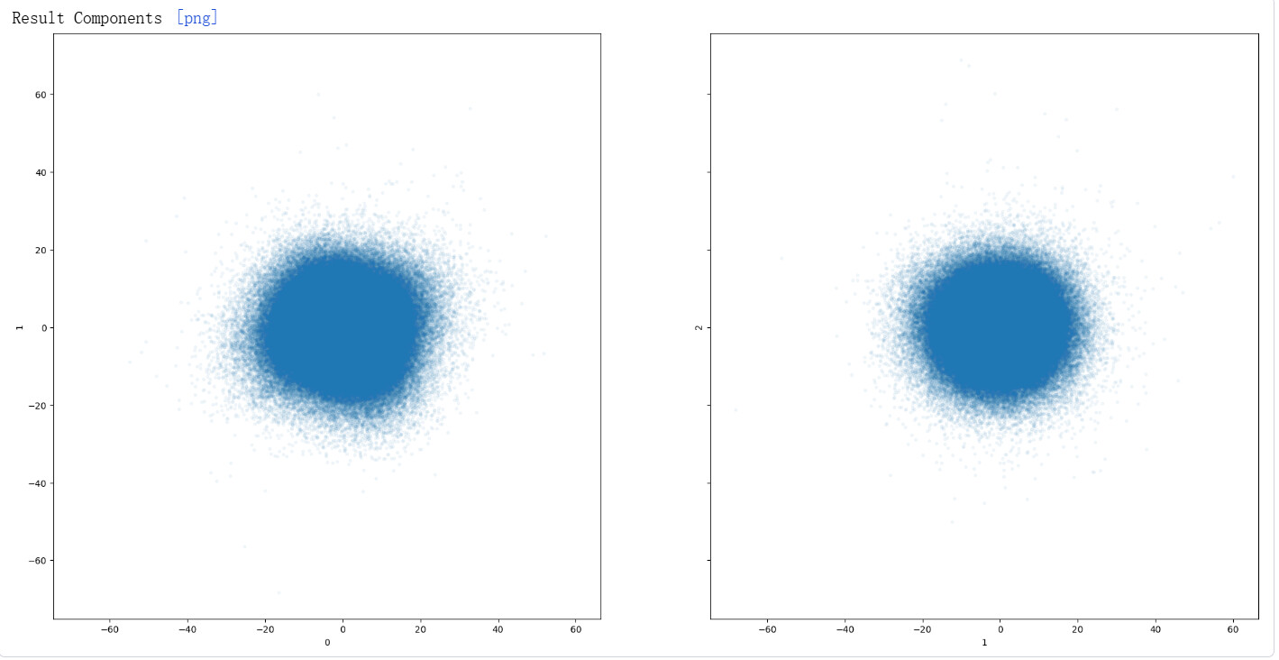
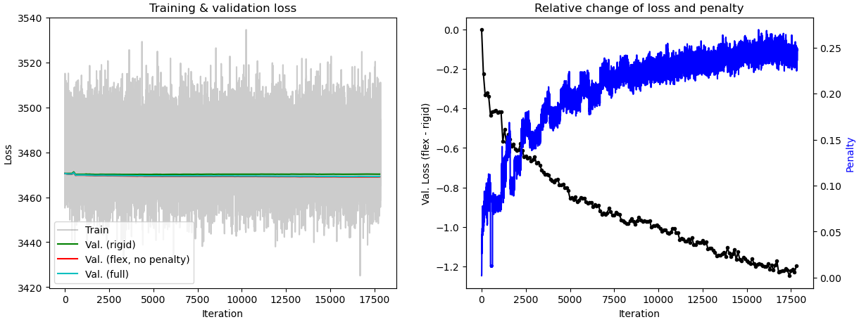
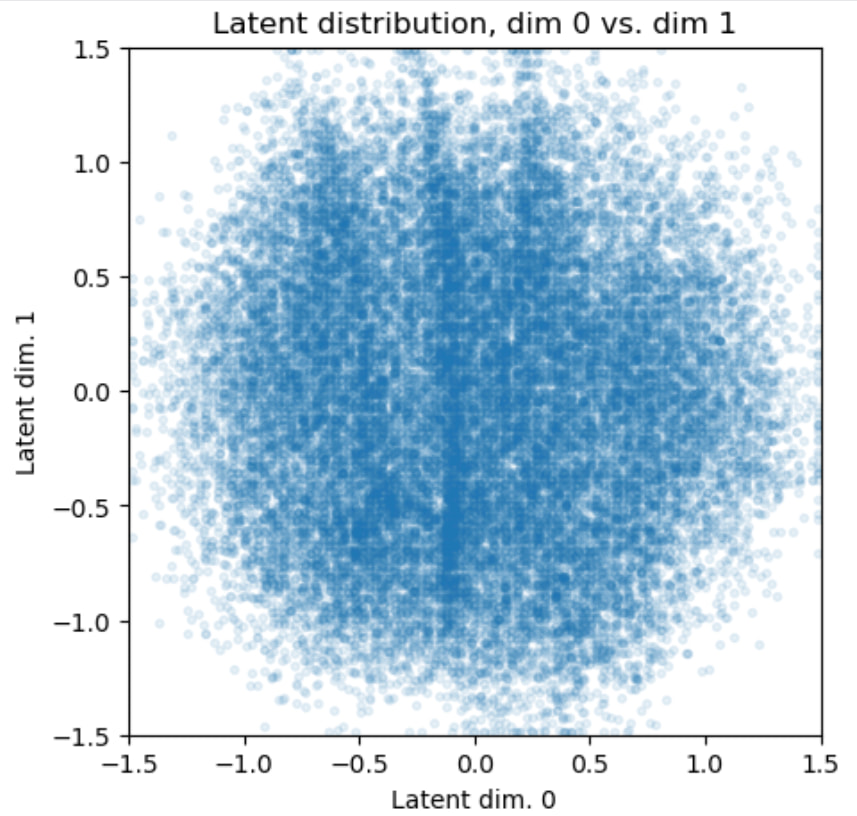
I’ve spotted elliptical component distributions in 3DVA and Display, so I ran 3D Flex Refine. I have already set rigidity to 0.05 and latent centering strength to 0.25. Even so, the validation curves Val (rigid), Val (flex) and Val (full) remain tightly overlapped. Does this mean the training has failed? Or should I evaluate map improvements after every reconstruction? Any advice would be greatly appreciated.
It’s hard to know whether your data work well with 3D Flex without seeing the results so far. Do you think you should show us an animation or images of the types of movenent 3DVA and 3D Flex each found?
My protein is an asymmetric complex consisting of an upper and a lower region, with an additional flexible domain on the side.
For 3DVA, I tested number of modes = 3 and 4, alongside filter resolution set to 8 Å and 10 Å.After multiple trials, The main motions I observed come from the lower complex region and the side domain. I focus on the lower region’s movement, as this part has the worst resolution in my map.
One thing puzzles me: the Result Components distribution in 3DVA looks nearly circular, but I can see the lower and side domains moving in 3DVA Display.
I also attempted local refinement, but the improvement was limited. It barely helped the poorly resolved region and the interfaces. Therefore I turned to 3D Flex to tackle the weak local density issue.
For 3D Flex training:Number of latent dims = 4; Latent centering strength = 0.1; I tested Rigidity values of 2 and 0.5 separately.
From the movie generated by flex, the lower section shows very little movement, and the side domain exhibits no visible motion at all. It seems the model has not captured meaningful conformational dynamics.
Could you give some advice on what might be causing this and which parameters or settings I should adjust?