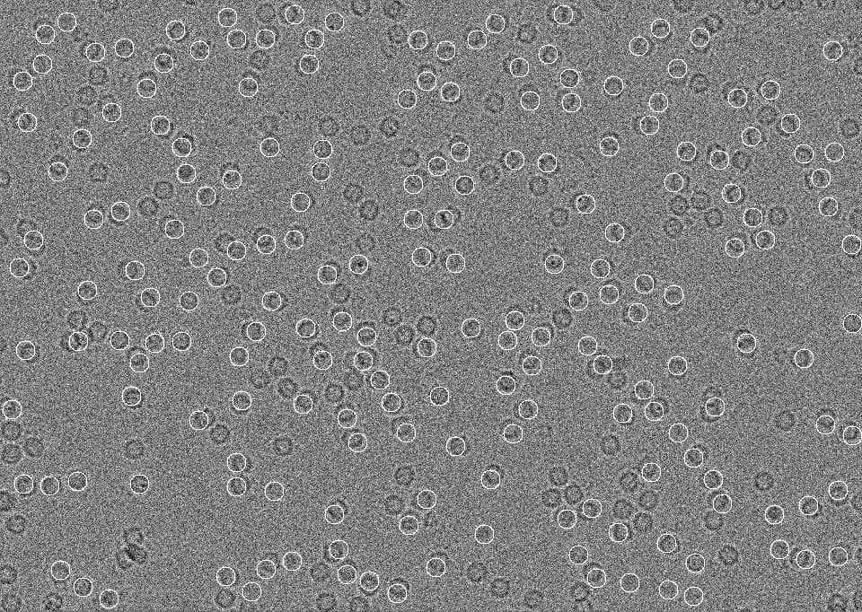
I’m reconstructing human apoferritin and got strange particle picking. For some reason most of the particles are cut off.
I’m not sure if this is because the microscope wasn’t aligned or a different problem that can be solved with the correct parameter.
To get my final reconstruction I adjusted the extraction box size and also expanded the expected diameter, but this isn’t a solution.
Here’s what the inspect particle picks job looked like:
Picking parameters would be useful to know for this.
For apoferritin, I usually use 90Å (min. diameter), 110Å (max. diameter) and ring blob. That has never failed to give nicely centred apoferritin picks (and yes, I know apoferritin is larger than that, but nevertheless…)
Are the picks unaligned because the microscope was unaligned or is there something else going on?
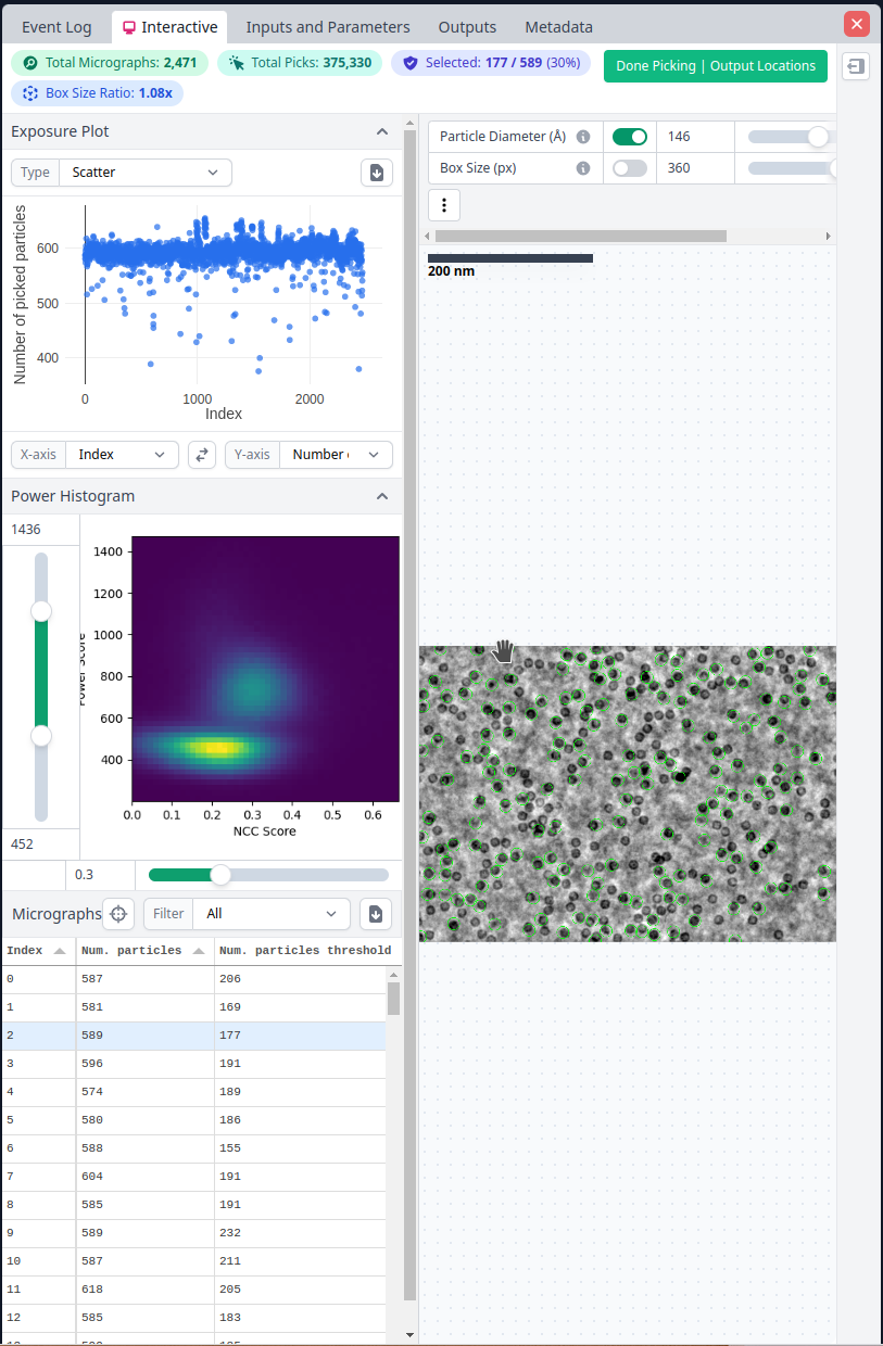
Picking Parameters:
Min diameter: 120 angstroms
Max diameter: 160 angstroms (typically I only put 140, but I got a bad 2D classification when I tried with 140)
Extraction box size: 360 pix (typically I would only use 280 when the max diameter is 140)
This is the NCC threshold and power histogram I chose:
280 px with your pixel size is about 135 Å and way to small for apoferritin. Even at 360 px your box is only about 1.33 times the particle diameter, which is on the small side and even more so if the picks are not well centered. Try a larger box, even if you’ve improved your picking.
Is there a standard for how much bigger the box size should be than your particle diameter? I was under the impression that the box size should match (mostly) the largest expected diameter.
The max/min diameter is very off for apoferritin - that will be the cause of the mis-centring I think.
Regarding box size, the “optimal” box size depends on a variety of factors. A decent general case is double your particle size (so apoferritin at 0.48 angpix would be ~540 pixel boxes). Due to signal delocalisation by the CTF, you’ll lose high frequency (resolution) information if the box is too tight. Higher defoci also need larger boxes due to the point spread function (PSF). Acceleration voltage also plays a role.
Apoferritin is incredibly forgiving for using non-optimal parameters and still getting good results (the high symmetry helps): our internal training dataset is optimal at 480 pixel boxes, but loses <3% of maximum resolution by cutting the box down to 400 pixels. A friend at another institute processed it with 360 pixel boxes (far too tight for my comfort) and reported a good result, though.
There is a nice examination of some of this in this manuscript: