I’m working on a protein (90kD) containing multiple domains connected by several long, flexible regions, and I’m trying the HR-HAIR strategy described by professor Oliver B. Clarke .
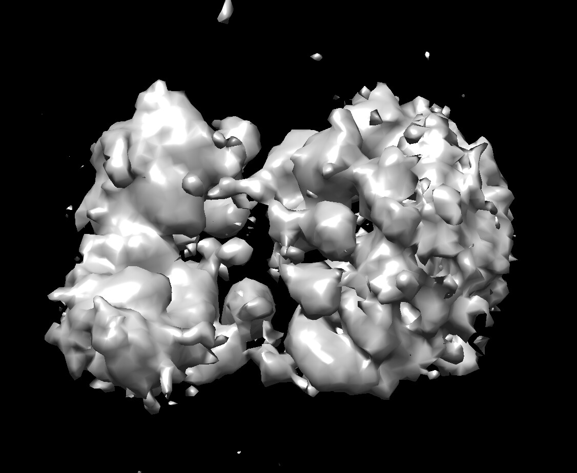
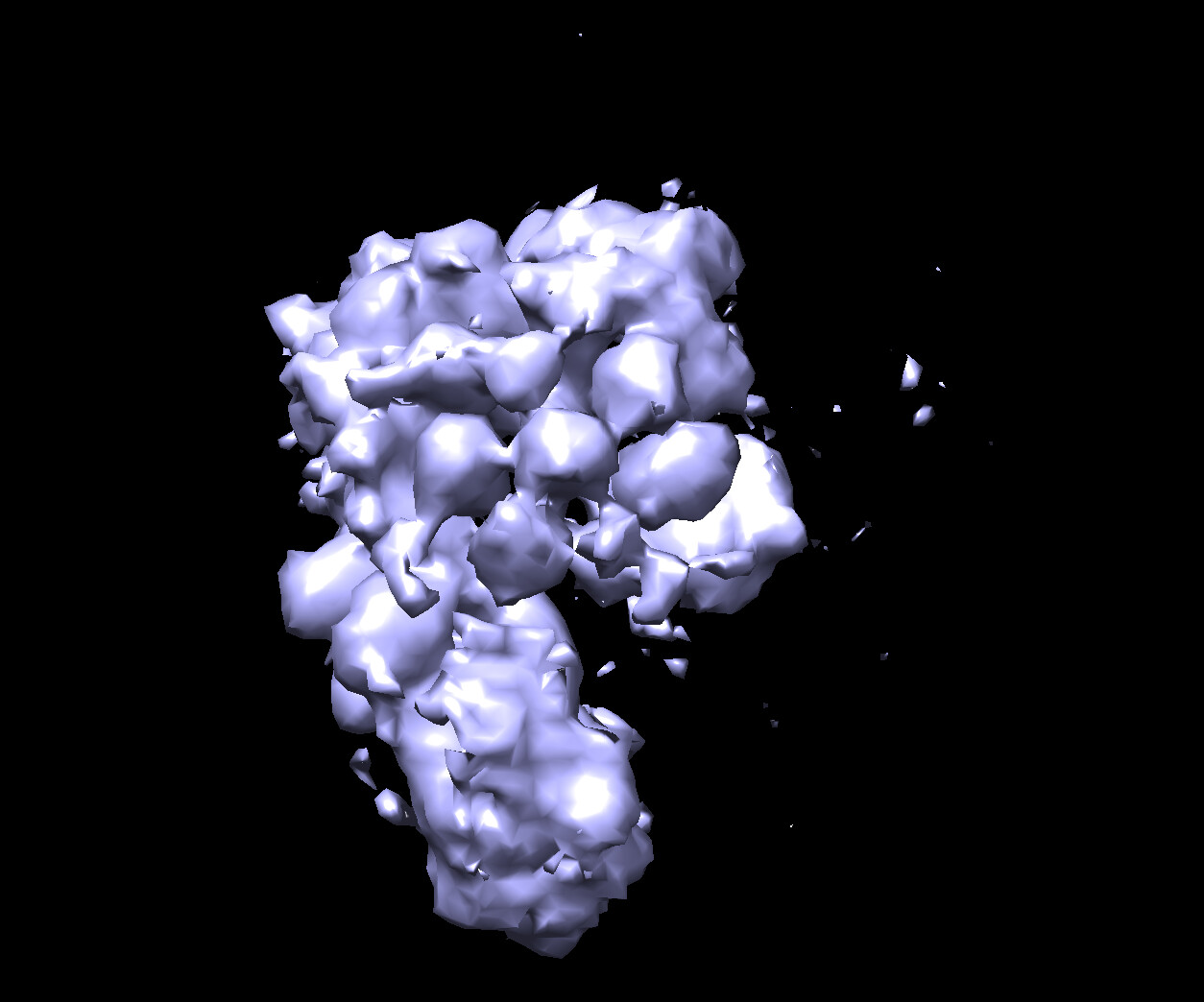
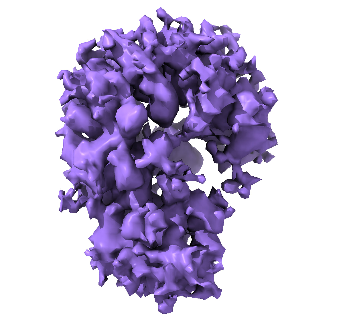
Using the parameters from the paper, the ab initio reconstruction shows poor secondary structure. Interestingly, at around 3,000 iterations, the reconstruction looks much better, with recognisable β-sheets. However, by around 4,800 iterations, it becomes worse again. Now it keeps going.
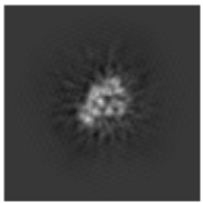
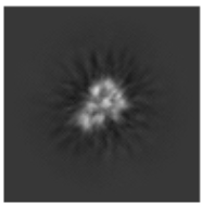
I also found that my 2D classification is much better with “Enforce non-negativity” enabled, whereas adding “Use clamp-solvent to solve 2D classes” together with “Enforce non-negativity” makes the classes considerably worse.
Has anyone encountered similar behaviour with highly flexible, multi-domain proteins? In particular, is it reasonable to use the ab initio reconstruction from an earlier iteration when it looks better, rather than the final reconstruction? Is it possible that the heterogeneity makes reconstruction difficult?
Any advice would be greatly appreciated. Thank you!
is the way to hell… unless you have some backfolding, you might have more success going back to the bench and making more rigid constructs. There are several strategies using binders for that kind of situation.
That said, if you want to try stuff on this dataset: try finding back folded classes and working with them only. This is still not a case for beginners because the protein is in the small side for cryoEM.
(I’m still curious about what the others have to say)
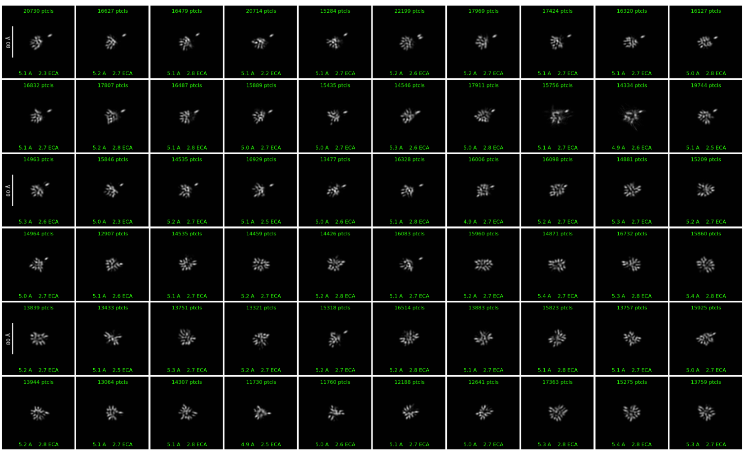
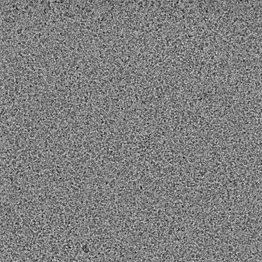
Are those really 2D classes of particles? I am a cryoEM beginner myself, but if I saw those 2D classes and they would become worse with iterations, I would think it is just noise. How does one know whether what they are looking at is particles vs. noise?
I agree with your concern. I only obtain these classes when I enable “enforce non-negativity.” I wonder whether enforcing non-negativity could result in the loss of some high-frequency signal or otherwise affect the 3D reconstruction.
I’m afraid your binders are still too small, unless they help locking the back folded state. Even with a large binder, if it allows movement of the other domains, it won’t help much (it should help with picking, though).
If you’re seeing better reconstructions at 3,000 iterations as opposed to 4,800, you can always ‘kill’ the job then ‘Mark as Complete’ and use the output volumes at that iteration. I’ve done that in the past when ab initio produces weird volumes after many iterations. This typically happened to me when I had preferred orientations.
Have you tried carrying these volumes through heterogeneous refinement yet?
Yes, I have tried heterogeneous refinement, but the result was not good enough—the resolution was around 8 Å. Now I intend to approach it from a few different steps:
Go back to the 2D classification, turn off “enforce non-negativity,” and select only the good classes based on the 2D classification results.
Manually separate the 2D classes according to the volumes I obtained previously, and reconstruct them separately.
Perform 3D classification and heterogeneous reconstruction, and try to obtain a “good” model with clear backbone density.
I have worked on and helped on some small flexible proteins so hope my insight can be helpful.
The 2D classes you currently have do not look the most detailed, though they are of a consistent size. Because of this it is probably too soon to go to 3D reconstruction.
Do the 2D classes look like protein with secondary structure when you don’t enforce non-negativity? For small proteins and ones with ordered domains it usually helps to turn off “Force max over poses/shifts”, increase the number of online-EM iterations to 40 or higher, and increase the batch size per class to 400 or higher. Sometimes using a soft circular mask can help as well if the micrograph is crowded.
Once you have 2D classes that do show protein features only then would going to 3D make sense. In my experience, before going straight to HR-HAIR, I have found using higher resolutions for the initial and maximum resolution can help. Maybe try 9 angstroms for the initial and 5 for the maximum. Also increasing the initial minibatch size to 300 and the final minibatch size to 1000 can help.
Compared with the transmembrane proteins I have processed before, these classes do indeed look like noise to me. Interestingly, after two rounds of 2D classification with “enforce non-negativity” turned on to obtain a relatively “good” particle set, which has high resolution value and reasonable ECA value, I performed one additional round of 2D classification with “enforce non-negativity” turned off. In this final round, I was able to obtain some classes with recognizable and reasonable 2D features.
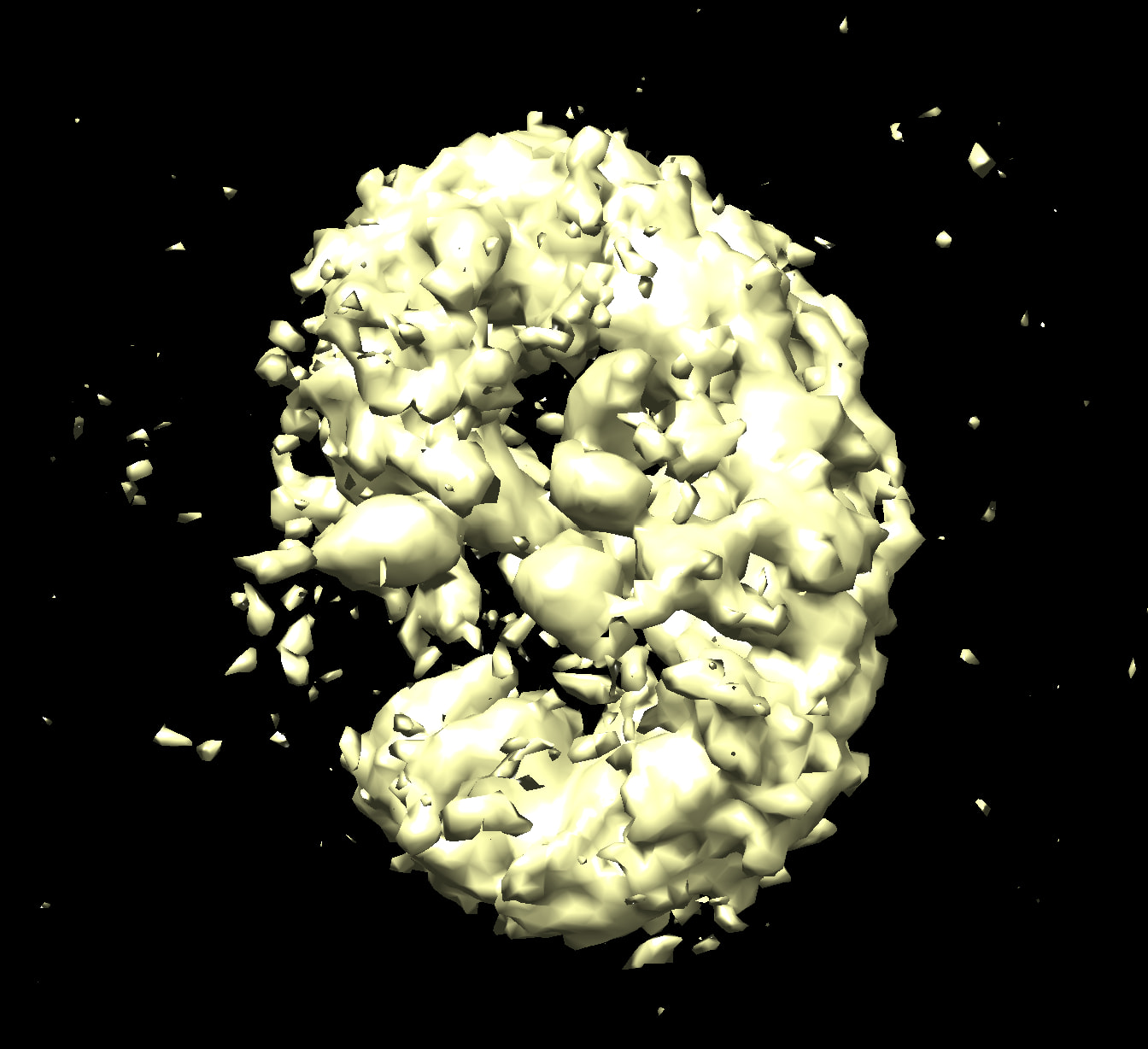
After 2D classification to exclude particles with obviously different parts of the protein, I rebuilt the ab initio model and performed 3D classification and heterogeneous refinement. This resulted in a better volume, as shown here. The reconstruction was optimized from the third model I posted previously (yellow).
But the volume is not good enough yet, and still needs further optimization.
After 2 rounds of 2D classification with “enforce non-negativity” turn on, additional round of 2D classification with “enforce non-negativity” turn off was performed and there are some good classes with 2D features, which look like the side view of the 3D ab initio model previously posted.
But as you can see, the features were not clear enough, I will try to turn off “Force max over poses/shifts” as you suggested in the 2D classification.
And thank you again for the advice about 3D reconstruction.
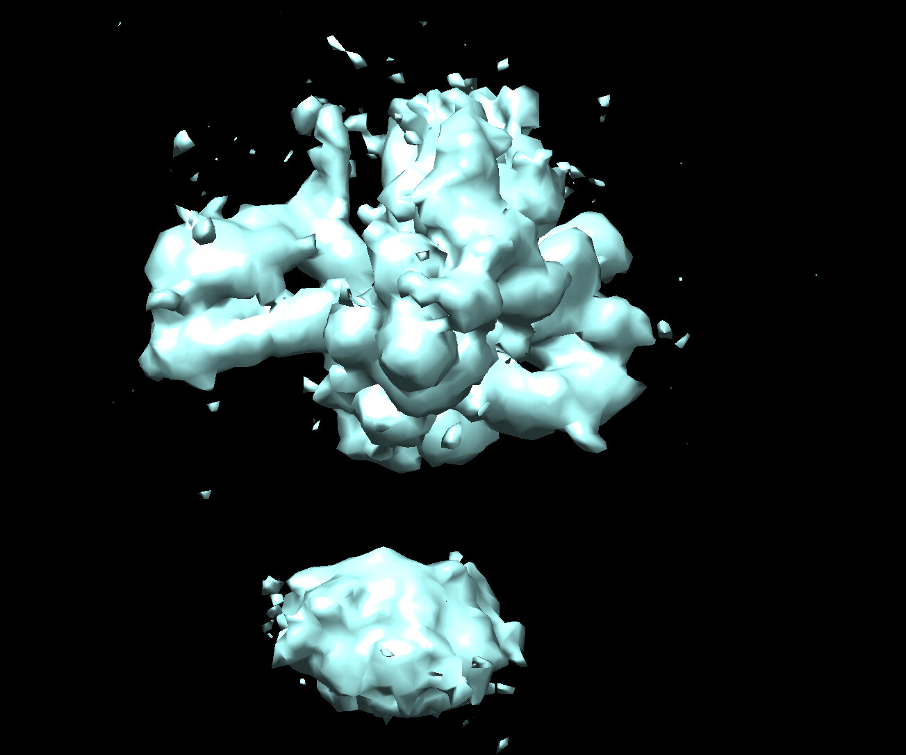
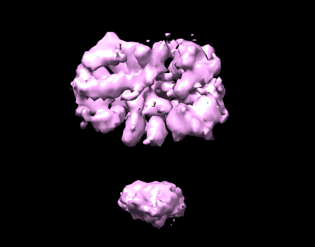
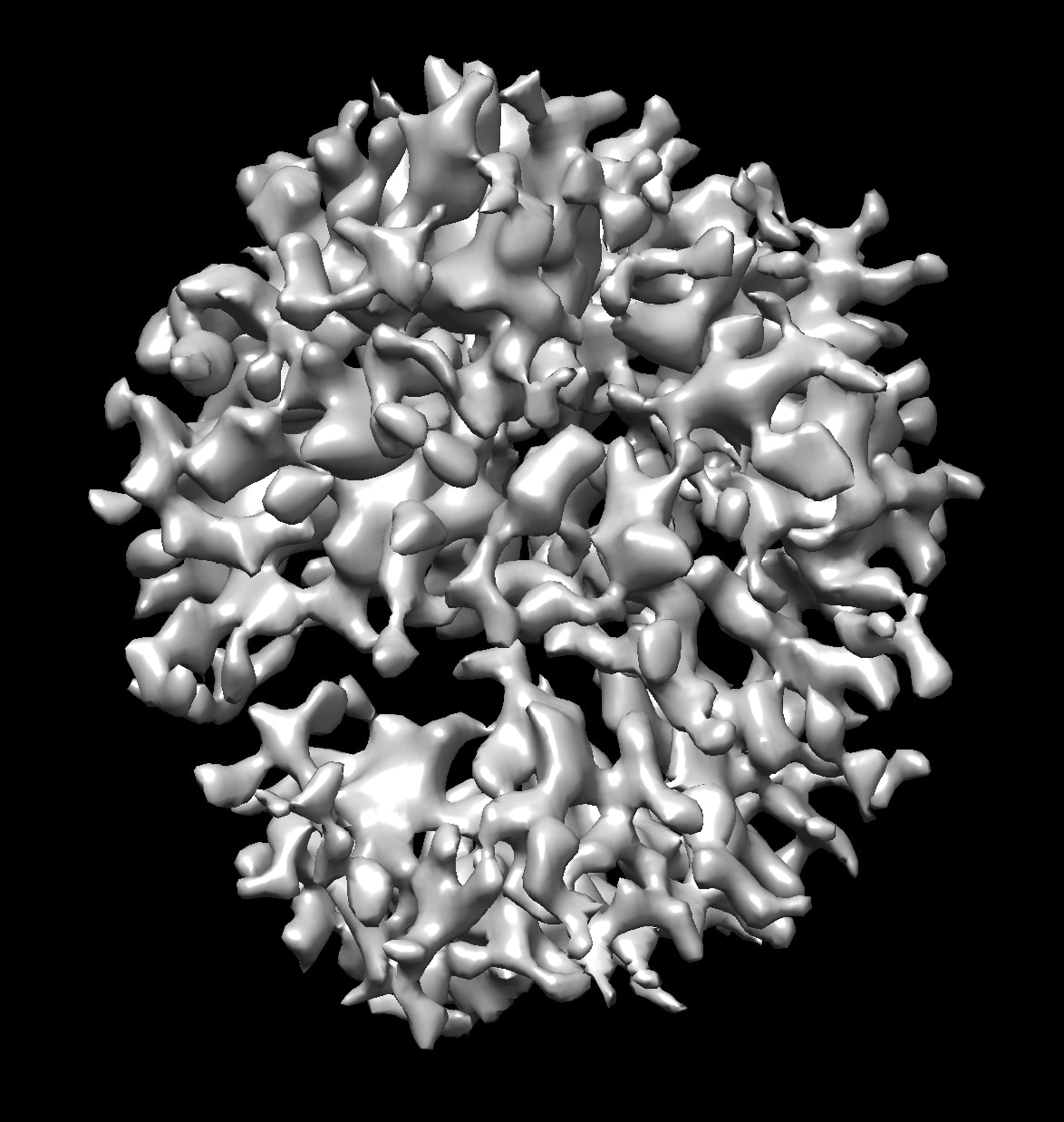
I got a better map,with an estimated resolution of 3.6Å, after repeating 2D classification and collecting only good classes. I will work on improving it further.
I still have questions about cases like mine ( protein adopt multiple conformations or had different parts) when performing HR-HAIR:
Would it help to use ab initio reconstruction with more classes (maybe 5-8) to distinguish reliable true classes? (in my case, I found reliable classes by processing two independent datasets collected at different times and reconstruct structures separately, then I got similar two). In addition, the resolution of ab initio reconstruction in my case was not as good as your published before, could it be because the particles from different conformations disturbed each other?
How should I recover all good particles which should belong to the true classes, but were assigned to the junk model during ab initio reconstruction? Is the particle assignment during ab initio reconstruction definitely correct? Should I perform 3D classification using the “true” class volume as a reference to rebalance or reassign the particles?
Hi Daniel, at least from this picture this does not look like a 3.6Å map I’m afraid. If you filter to lower resolution, can you see evidence of secondary structure? I would always suggest paying more attention to what the map looks like than to the estimated resolution.
I agree with Oli. Unfortunately, it doesn’t look as if there are secondary structure features in your new map. Is the map the expected size of your protein or of a similar size to any of the expected folded domains?