Hi everyone,
I’m a beginner in cryo-EM and currently working with my first dataset.
I’m studying curved protein filaments that form under acidic conditions (pH 2.3). The sample was collected from the pellet fraction after ultracentrifugation and resuspended in the same acidic buffer for purification and concentration. I used this sample to prepare grids with a Vitrobot using Quantifoil Cu R 1.2/1.3 grids. Data collection was performed on a JEOL CRYOARM 200 (200 kV) with a K3 camera in super-resolution mode, and 10,000 movie frames were collected.
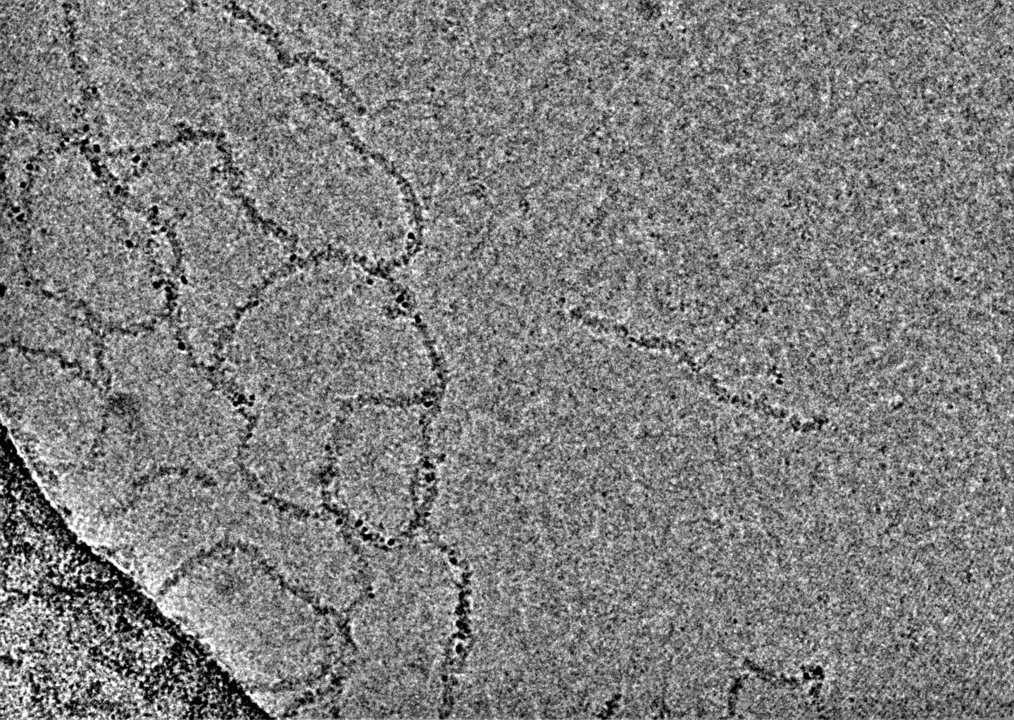
Following the helical reconstruction tutorial in cryoSPARC, I performed Patch Motion Correction and Patch CTF Estimation, and then removed micrographs with CTF fits worse than 6 Å using Manually Curate Exposures (Figure 1).
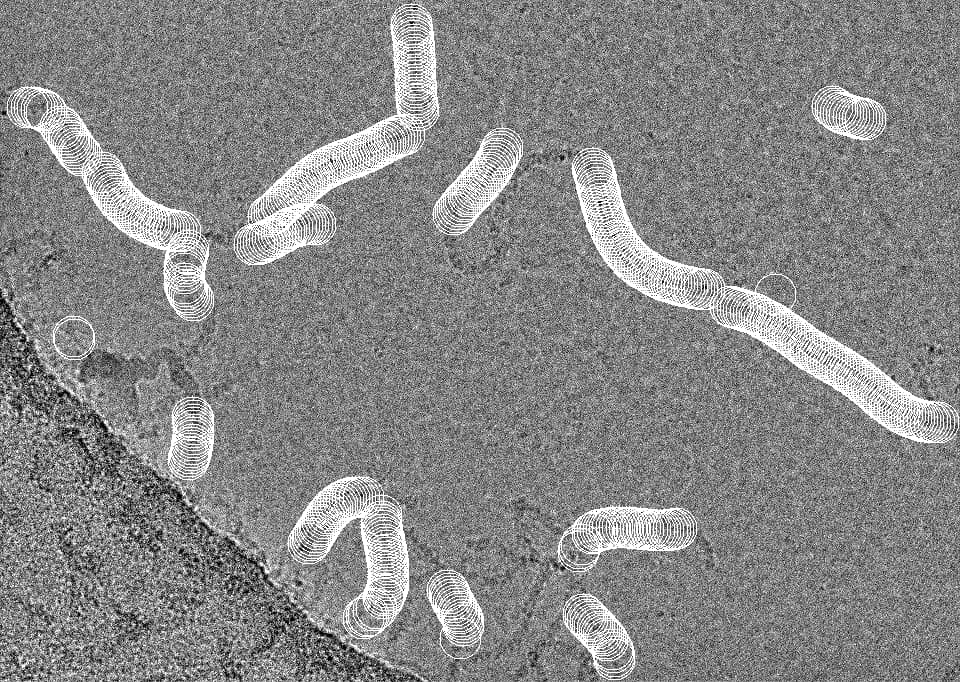
Next, I performed manual picking and created 2D templates for Filament Tracer, which I used to detect filaments on 500 curated micrographs, followed by Inspect Picks for curation.
Filament Tracer settings:
・Filament diameter (Å): 70
・Separation distance between segments (diameters): 0.25
・Number of micrographs to process: 500
Inspect Picks thresholds:
・NCC score > 0.280
・Local power > 1287.000 and > 2374.000
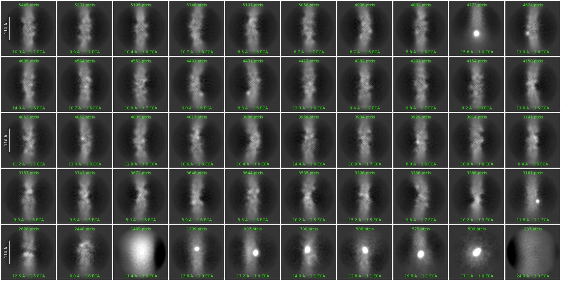
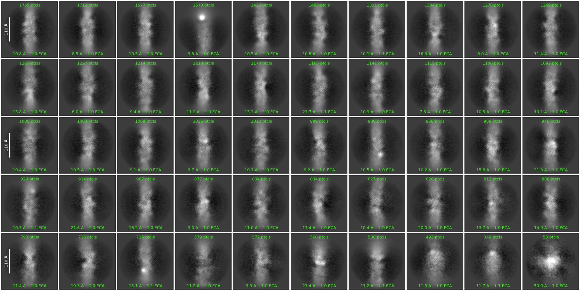
I then extracted the particles using Extract from Micrographs (box size: 300 px) and ran 2D Classification. However, the resulting classes were very low resolution and often showed spiral-like striations in the center (Figure 3). Since the filaments are inherently flexible, I tried removing the highly curved classes and re-running 2D classification, but the results remained low resolution (Figure 4).
1st and 2nd 2D Classification settings:
Batchsize per class: 400
Here are my questions:
Given the high flexibility of these filaments, I’m wondering if the standard helical reconstruction pipeline may not be suitable for this dataset. I’ve tried several 2D classification cleaning strategies suggested in previous forum discussions—for example, multiple ab initio classifications followed by 2D classification, or heterogeneous refinement followed by 2D classification, changing the extraction box size, etc. But in all cases, very few particles remain after classification, and the resulting ab initio models are not well-resolved. Do you have any advice or ideas for modifying the workflow to better handle such flexible filaments?
The filaments I’m working with are not typical amyloids and seem to have irregular internal structure and high flexibility. Do you think a strategy that involves collecting significantly more data, and then isolating a subset of more structurally homogeneous particles from a large population, makes sense in order to eventually obtain clearer 2D classes or a 3D model?
Thank you very much for any suggestions or insights!
We discuss in this paper how an asymmetric reconstruction for the Vibrio T4P yielded higher resolution than the helical reconstruction, due to the flexibility.
Thank you very much for sharing your valuable paper. Even in the absence of clear 2D classes, proceeding with 3D classification may indeed be a viable approach. I will also consider the workflow of ab initio reconstruction followed by heterogeneous refinement. I sincerely appreciate your guidance.
I am also working amyloid-related premature fibrils like protofibrils. The samples I deal with look like yours too. In my case, after the 2D classification, a periodic 4.8 angstrom-spaced ladder suggesting a beta-sheet repeat was observed at the center of a filament structure. However, no 3D reconstruction worked to obtain detail structure.
In my case, first I tried 2D classification using a 1/4 downsized particles, and not obvious signature was observed. Then I tried without downsizing, obtained the beta-sheet repeat structure.
So my simple question is if you performed the 2D classification using non-binned particles, i.e. without downsizing the images.
Though my comment might not work on solving your problem, I hope it will help to solve your problem in any ways.
I feel happy to know people working on similar samples!
Have you tried 2D with a smaller mask - just covering maybe 1.5 filament widths? this might help reduce the influence of filament flexibility a bit (and you can then proceed with ab initio as per @egelman’s comment, using the best 2D classes).
Thank you very much for your helpful advice.
I adjusted the mask size to cover about 1.5 filament widths and re-ran the 2D classification. As a result, several 2D classes appeared that showed distinct and detailed structural features. I selected those classes and performed ab-initio reconstruction followed by heterogeneous refinement using multiple classes.
However, I was not able to obtain any interpretable 3D volumes. I suspect this is because the region of interest is too small. Therefore, I am now trying to follow the approach described in your recent bioRxiv preprint, “High-resolution ab initio reconstruction enables cryo-EM structure determination of small particles”, using HR-HAIR as a reference.
Since my sample is quite flexible, HR-HAIR might not be directly applicable—but I’m giving it a try. I would greatly appreciate any further advice you might have. Thank you again for all the valuable suggestions from everyone here.