The job ‘extract from micrographs’ will extract particles from the micrographs. I have open the file .mrc which stores the particles. However, none of them contains observable particles, though I can get the true 3D structures.
Plus, the particles are 440*440, and each image looks like this:
I want to use the extracted particles for other tasks. Do you have any ideas? I am looking forward to your reply.
Regards,
Benjamin
Hi @benjamin_shi, can you please explain your workflow a bit more?
- Which type of particle picking job(s) did you use?
- Were you able to use Inspect Picks to visualize and adjust the picks before extracting them?
- Were you able to run 2D classification on the extracted particles after Extract from Micrographs?
Hi, spunjani
@spunjani
I am just following the T20S tutorial. I reconstructed the structure pretty well. However, I just want to know how to extract the particles to individual files e.g. .png/.tif.
(1) I did manual picking followed by template picking
(2) YES, I can inspect picks before extracting them. The images shown in cryoSPARC before and after extracting are both quite good.
(3) YES, I can run 2D classification and finally do ab initio reconstruction and refinement of T20S correctly.
Everything is good, and I can perform the 3D reconstruction correctly.
However, when I examine the .mrc images generated at each step (e.g. patch motion correction, particle picking, extract of particles), I can not observe any particle in these noising images (only the class average is recognizable). I use imageJ or python codes to open these image stacks and convert them into image. Have you done some filtering to make the particle visible as shown in cryoSPARC?
All in all, the images shown in UI and preserved in the folder are not quite the same:
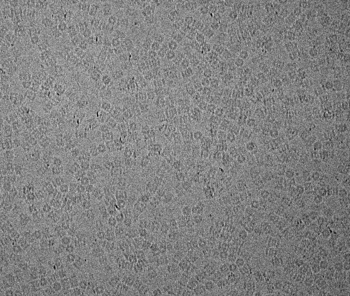
Original image and the image shown in the webapp :
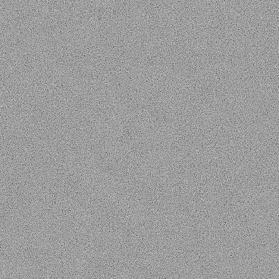
The image in the folder of the job “Patch motion correction”.mrc
Ben
Hi @benjamin_shi,
Good questions. What you are seeing when you open the .mrc stacks in imageJ or python and display them “raw” is the actual cryo-EM data that was collected. Yes, it really is that noisy and you can’t see anything - this is expected. The signal-to-noise ratio of raw data is typically worse that 0.01.
It is only by low-pass filtering the images and downsampling that the particles start to become visible in the images. We always display filtered images in cryoSPARC. In the interactive jobs, there is a slider that controls the corner frequency of the filter so you can play with that and see how the image changes as more of the high-res noise (and signal) is removed.
Hope this helps!
Hi, @apunjani
Sorry but it doesn’t make too much sense. I mean, I can perceive the particles in the original experimental images, but I cannot observe the particles in the micrographs where your extracted particles.
Ben