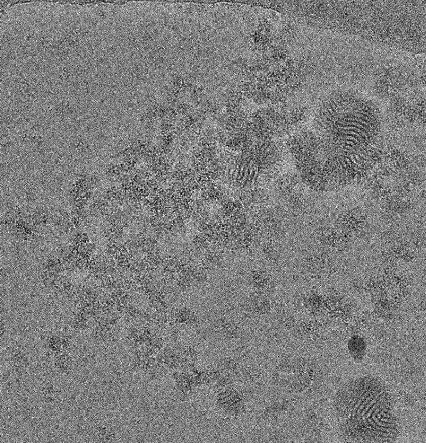
As the subject line indicates, I have a peculiar artefact in my micrographs. I see these on gold and continuous carbon grids. My sample is a membrane protein extracted in digitonin and vitrified in LMNG or GDN. I see this artefact irrespective of grid type, detergent used, or changes in buffer conditions. I have never seen these before, nor has anyone else I’ve spoken to. Does anyone have an idea what this could be?
This sure is an interesting case… I’m curious to see what the FFT looks like in the affected images.
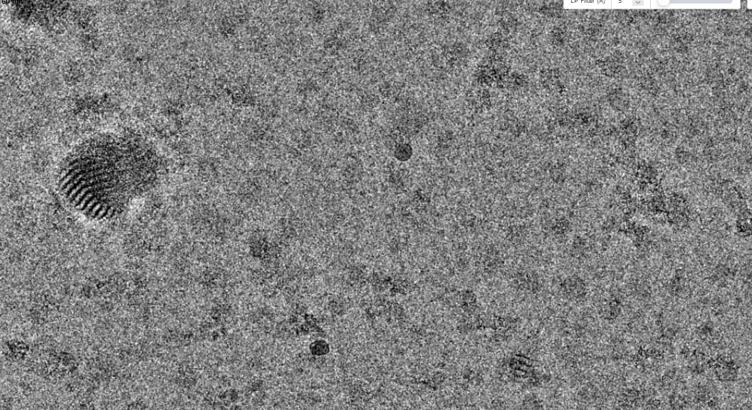
I’m thinking that this is either a problem with the ice (i.e. partial crystallization of some sort?) or aggregation/assembly of your protein sample. That said, I believe you have
crystalline ice in the second image you’ve posted (lattice lines visible at the top left corner) which may or may not be related to the problem.
If the problem is indeed reproducible under various buffer/detergent conditions, then it’s probably more than likely related to your specific protein of interest. Judging from image #1, your protein does have a tendency to aggregate, and it almost seems like the fossil artifacts are “growing out” of the aggregated protein mass.
Is it possible that your membrane protein is capable of forming multi-layered complexes (like in the case of aquaporin) that upon aggregation may lead to the striated “artifacts”?
We have seen similar with CHAPS/cholate/deoxycholate - fibrils, sheets etc.
I bet if you pick all over them and run Class2D you will see some quite ordered classes!
Although the fact that it doesn’t vary with detergent is surprising - I haven’t seen this with digitonin personally. Then again, digitonin is so heterogeneous that it could be batch to batch variation…