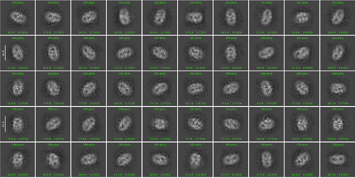
The 2D image shows that the shape of these particles is as expected for my protein. But is the poor 2D result because the particles come from a thicker part of the ice, or is the particle itself a poor signal? Will it improve if I increase the number of iterations?Could it just be empty detergent micelle?
Hi, first thing I noticed is the low particle counts per class. Only around 300 in each class, this may not be enough to form a high-res 2D classes. In the first instance I would suggest to increase the number of micrographs/particles to see if you get better 2D classes. If you are reaching around 10000 particles per class and still not getting good 2D classes, that would be the indication of something wrong with the sample/ice.
Secondly, this looks like membrane protein/micelle? This further complicates things as there could be some empty micelles there which blurs out the 2D.
