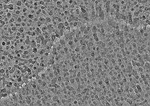
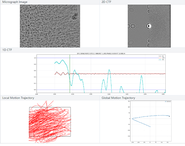
Excuse me, has anyone ever seen this kind of micrograph ? The particles shown are not protein samples. Is this caused by glycerol or solution contamination? Or is it caused by freezing or transferring? Thank you

First image is very bad ice (sometimes called leopard ice if I’m remembering my cryo-EM slang correctly) with a microfracture straight through it. Second is same ice condition, with the right hand side also being extremely drifty. It either picked up a lot of contamination during transfer, or partially liquified and re-froze at some point before imaging.
1 Like
As rbs_sci said, this is leopard ice seen usually when the grid thaws and refreezes. There maybe particles in these images but if majority of your grid looks like this then it is not worth collecting/processing.
