Hi everyone,
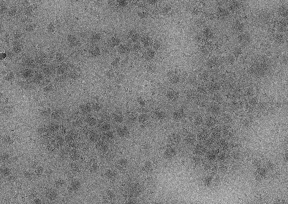
Has anyone had experience with grids coated with 2 nm carbon film? My sample is ribosomes, but the particles appear quite blurry on these grids and the structural details are not clearly resolvable.
Hi everyone,
Has anyone had experience with grids coated with 2 nm carbon film? My sample is ribosomes, but the particles appear quite blurry on these grids and the structural details are not clearly resolvable.
Good day Hai. Have you tried a range of blotting conditions that might vary the ice thickness, and does this happen across all conditions, and all areas of the grids? It’s possible we’re looking at one of the extremes, such as much too thin ice.
Hi @drichman thank you for your reply
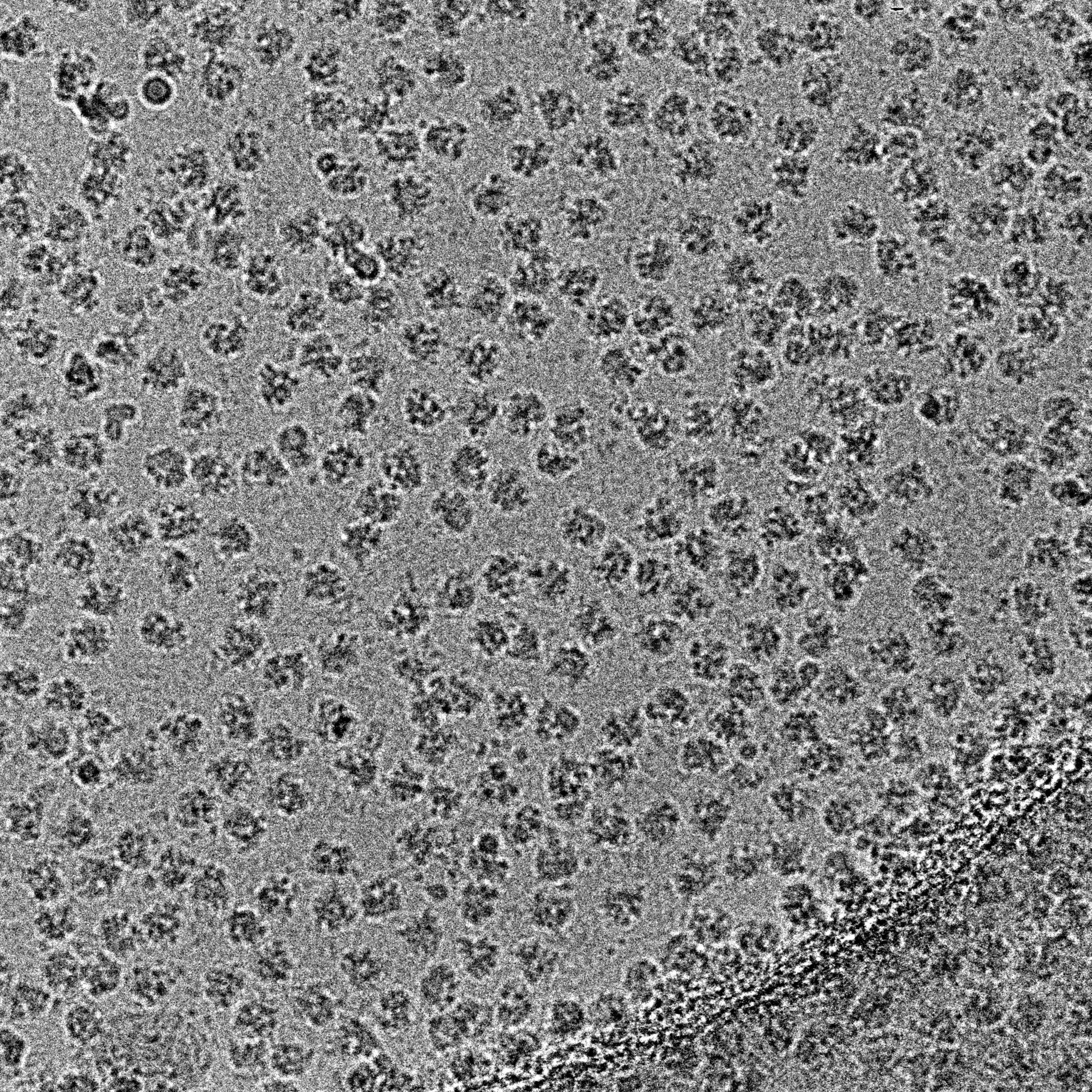
I will try adjusting the cryo-EM sample preparation conditions. Additionally, shown below is how the sample appears on 1.2/1.3 grids with the exact same preparation protocol.
Can you tell us the acceleration voltage, magnification, defocus, camera and total dose of the exposures? Otherwise, it is difficult to judge the contrast. I guess the first micrograph is “data collection” type of micrograph on a Krios with perhaps 0.8Å/pixel, 30-40 e/Å2 and perhaps -1 um defocus and that the lower one is Glacios with 1.2Å/pixel, 80 e/Å2 and -2 um defocus? Those differences will give very different (low resolution) contrast.
Thanks for your reply.
The first micrograph was acquired on a CRYO ARM 300 electron microscope (JEOL, Japan). The data collection parameters are as follows:
Pixel size: 0.475 Å (super-resolution mode)
Accelerating voltage: 300 kV
Spherical aberration: 2.7 mm
Total exposure dose: 50 e⁻/Ų
Defocus range: -1 to -2.5 μm
The second image was captured during routine sample screening on a 200 kV microscope, and I cannot recall the exact acquisition parameters.
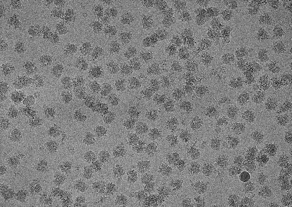
In fact, we previously obtained fairly sharp images using +2 nm carbon film copper grids under the exact same imaging conditions as the first micrograph. Those reference images are shown below.
Ok, the other example you show looks good, but without knowing the defocus for the exact micrograph in the original post, it is difficult to say if this is due to thickness variations of the continuous carbon, in thickness of the ice or just defocus difference.