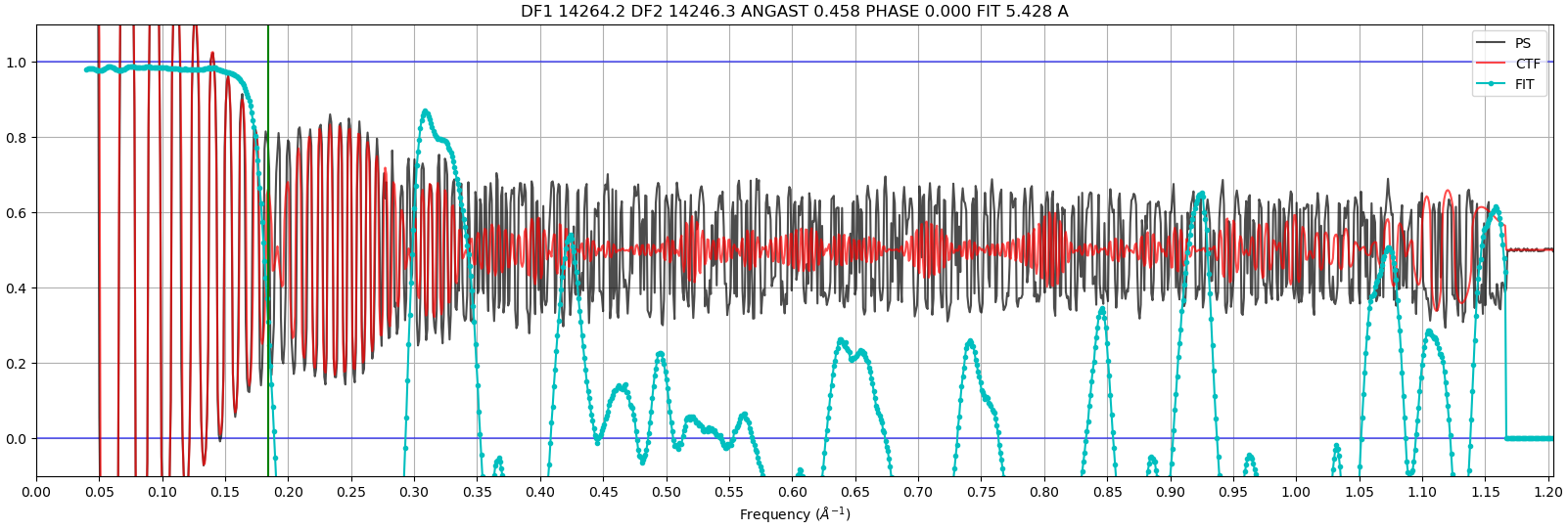
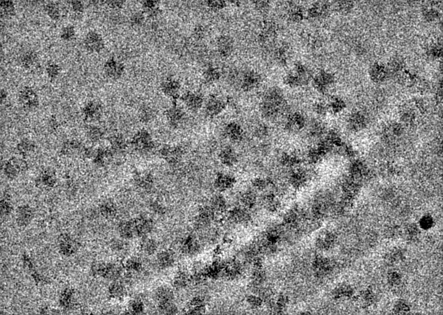
I am processing cryo-EM data to obtain ribosome maps, but I am having trouble generating 2D classes. I have attached my CTF fit image and the average micrograph.
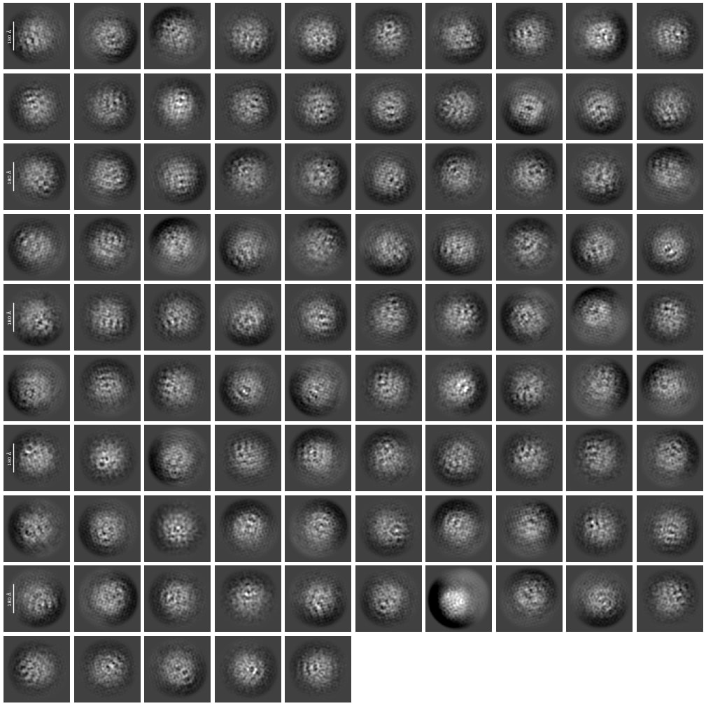
The micrographs clearly show the presence of ribosomes, although there are also some fringe-like artifacts. However, when I perform 2D classification, the resulting classes consist entirely of these fringes rather than ribosome particles.
I was wondering if anyone has encountered a similar issue or has any recommendations on how to fix this problem. Any suggestions would be greatly appreciated. Thank you!
Looks like a few different issues going on here. One is that your CTF fits are very low res, that often suggests that the ice is extremely thick. I would think that would be the primary reason for what you are seeing.
The other issue is that the image you shared has not been properly gain corrected. Check to see if you supplied the gain ref or if it requires rotation/flipping. That said, you should be able to get good 2D classes even you didn’t supply any gain ref. I think the data/ice quality is the bigger issue here.
Agree with all of this, and I would also check your box size - a mask ~200Å seems too small for a ribosome? (though I am not a ribo guy, so take that with a grain of salt)